The Structure of the Heart (AQA A-Level Biology): Revision Notes
The Structure of the Heart
The heart is a muscular organ located behind the breastbone in the chest cavity. It operates continuously throughout an organism's life, functioning as a vital pump that circulates blood around the body.
The human heart consists of two separate pumps positioned side by side. The left pump handles oxygenated blood returning from the lungs, while the right pump deals with deoxygenated blood returning from the body tissues. This dual-pump system prevents the mixing of oxygenated and deoxygenated blood.
The separation of oxygenated and deoxygenated blood is crucial for efficient circulation. This dual-pump design ensures that oxygen-rich blood from the lungs doesn't mix with oxygen-poor blood returning from body tissues, maintaining optimal oxygen delivery throughout the body.
Heart chambers
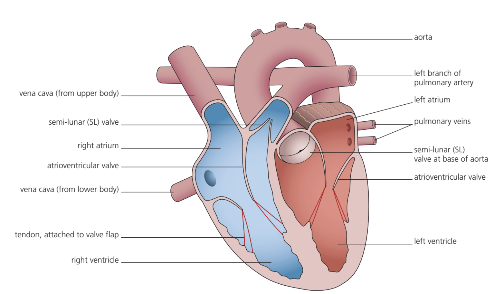
Each pump contains two chambers, giving the heart a total of four chambers:
Atria (singular: atrium)
The atria are the upper chambers of the heart. They have thin, elastic walls that allow them to stretch as blood collects within them. The atria serve as collecting chambers that receive blood returning to the heart.
- Right atrium: Receives deoxygenated blood from the body
- Left atrium: Receives oxygenated blood from the lungs
Ventricles
The ventricles are the lower chambers with much thicker muscular walls compared to the atria. This thick muscle layer enables them to contract forcefully, generating sufficient pressure to pump blood over considerable distances.
- Right ventricle: Has a thinner muscular wall as it only needs to pump blood to the nearby lungs
- Left ventricle: Has the thickest muscular wall since it must generate enough pressure to pump blood throughout the entire body
The difference in wall thickness reflects the different distances blood must travel and the pressure required for effective circulation. The left ventricle's thick walls are essential for generating the high pressure needed for systemic circulation.
Heart valves
Atrioventricular valves are located between each atrium and its corresponding ventricle. These valves prevent the backflow of blood into the atria when the ventricles contract.
- Left atrioventricular valve (bicuspid valve): Located between the left atrium and left ventricle
- Right atrioventricular valve (tricuspid valve): Located between the right atrium and right ventricle
Both sides of the heart contract together - first both atria contract simultaneously, then both ventricles contract together, ensuring coordinated pumping action.
Major blood vessels
Four large blood vessels connect to the heart chambers, carrying blood to and from the heart:
Vessels connected to the left side
- Aorta: Connected to the left ventricle, carries oxygenated blood to all body parts except the lungs
- Pulmonary vein: Connected to the left atrium, brings oxygenated blood back from the lungs (unusual for a vein to carry oxygenated blood)
Vessels connected to the right side
- Vena cava: Connected to the right atrium, brings deoxygenated blood back from body tissues
- Pulmonary artery: Connected to the right ventricle, carries deoxygenated blood to the lungs (unusual for an artery to carry deoxygenated blood)
Memory Aid: Atria link to Veins, and Arteries link to Ventricles
This simple mnemonic helps remember the connections between heart chambers and major blood vessels. Notice that the pulmonary vessels are exceptions to the typical rule about what type of blood arteries and veins carry.
Coronary circulation
Although oxygenated blood passes through the left side of the heart, the heart muscle itself does not obtain oxygen from this blood. Instead, the heart muscle receives its own dedicated blood supply through coronary arteries, which branch off from the aorta shortly after it leaves the heart.
Critical Concept: Blockage of coronary arteries can lead to myocardial infarction (heart attack), where part of the heart muscle becomes deprived of oxygen and may die. This occurs because the muscle cells in the affected area cannot respire aerobically and eventually die.
Understanding coronary circulation is essential because heart attacks result from problems with this separate blood supply system, not from issues with blood flowing through the heart chambers.
Why two separate pumps are necessary
The dual-pump system exists because blood must pass through tiny capillaries in the lungs to allow efficient gas exchange. This passage through narrow vessels causes a significant drop in blood pressure. If the same blood continued directly to the body tissues at this low pressure, circulation would be very slow and inefficient.
By returning the blood to the heart after passing through the lungs, the pressure can be increased again before distribution to the rest of the body. This ensures rapid and efficient circulation of oxygenated blood to all body tissues while maintaining the essential separation of oxygenated and deoxygenated blood.
Key Points to Remember:
- The heart consists of two separate pumps, each with an atrium (thin-walled) and ventricle (thick-walled)
- Atrioventricular valves prevent backflow when ventricles contract
- The left ventricle has the thickest wall as it pumps blood around the entire body
- Four major vessels connect to the heart: aorta, vena cava, pulmonary artery, and pulmonary vein
- The heart muscle receives oxygen through its own coronary arteries, not from blood passing through its chambers