Wave-particle duality (AQA A-Level Physics): Revision Notes
12.2.6 Electron microscopes
Resolving Power of Electron Microscopes
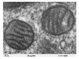
The resolving power of a microscope is its capability to differentiate between two closely positioned structures. This ability depends on the wavelength of the illumination source. In electron microscopes, the wavelength of an electron beam is significantly shorter than that of visible light, which gives electron microscopes a much higher resolving power compared to optical (light) microscopes. This makes electron microscopes ideal for observing extremely small structures, such as cell organelles, with high clarity.
The resolving power of the microscope improves as the wavelength of the electrons decreases, which can be achieved by increasing the accelerating voltage in the electron microscope, as this reduces the wavelength according to de Broglie's equation:
Where:
- is Planck's constant,
- is the mass of an electron, and
- is the velocity of the electrons.
Types of Electron Microscopes
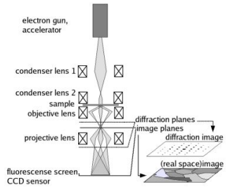
- Transmission Electron Microscope (TEM)
-
In a TEM, electrons are accelerated by an electron gun and passed through a series of magnetic lenses before reaching an extremely thin sample.
-
As the electrons travel through the sample, they experience minimal slowing, which maintains their short wavelength and thus the high resolving power.
-
The magnetic lenses serve various functions:
-
Condenser Lens: Directs and focuses the electron beam into a wide, parallel form that is aimed at the sample.
-
Objective Lens: Creates a magnified image of the sample above it.
-
Projector Lens: Further magnifies the image from the objective lens and projects it onto a fluorescent screen for viewing. Factors Affecting TEM Resolving Power:
-
Sample Thickness: Thicker samples slow down electrons, causing increased wavelength and reduced resolution.
-
Electron Speed Variation: Differences in electron speeds (due to energy loss in thermionic emission and collisions) lead to varied wavelengths, causing image blurring. Calculating Voltage for Atomic Resolution:
To resolve atomic-scale structures (around 0.1 nm), we can calculate the required accelerating voltage using de Broglie's wavelength formula. For example:
Rearranging for (accelerating voltage) provides the necessary potential to achieve atomic resolution.
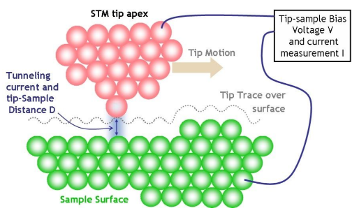
- Scanning Tunnelling Microscope (STM)
- An STM uses quantum tunnelling of electrons to create an image of a surface. This method relies on the wave nature of electrons: electrons can move across barriers (such as physical or potential gaps) if they are small enough.
- A very fine-tipped probe moves across the surface, kept at a constant potential. The tunnelling current (electron flow across the gap) varies with the gap size between the probe and the surface. This current can be measured and used to map the surface's structure.
Modes of Operation:
-
Constant Height Mode: the probe remains at a fixed height, and variations in tunnelling current create the image.
-
Constant Current Mode: the tunnelling current is kept constant by adjusting the probe's height, and this height variation generates the image. Image Formation:
-
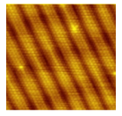
By keeping track of the probe's movements and the tunnelling current, STMs can achieve atomic resolution on conductive surfaces, mapping out the arrangement of atoms.