Gas Exchange System Tissues and Components (OCR A-Level Biology A): Revision Notes
Gas Exchange System Tissues and Components
The gas exchange system comprises multiple organs constructed from specialized tissues. Each tissue has distinct structural characteristics matched to its specific role. Understanding these individual components and how they combine within different airways is essential for appreciating how the respiratory system functions efficiently.
Tissue types in the gas exchange system
Cartilage
Cartilage is a type of connective tissue that provides structural support and reinforcement to airways. It consists of cells embedded within an extracellular matrix composed of mucopolysaccharides — complex polysaccharides containing amino groups. This tissue resists both tension and compression forces, offering flexibility superior to bone while maintaining adequate rigidity.
Within the respiratory system, cartilage prevents the collapse of large-diameter airways with thin walls. Without this cartilaginous support, pressure differences during breathing would cause these tubes to collapse inward, obstructing airflow.
The cartilage does not form continuous rings but appears at intervals, allowing the airways to retain some flexibility. This arrangement balances the need for structural support with the requirement for movement during breathing.
Ciliated epithelium
Epithelium refers to any cellular layer that forms a covering or lining on body surfaces. Ciliated epithelium is distinguished by the presence of cilia — hair-like projections on the cell surface.
Airways are continuously coated with a layer of mucus that traps dust particles and bacteria entering with inhaled air. The cilia beat in coordinated waves, creating a constant upward movement of this mucus layer toward the top of the trachea. Once the mucus reaches the oesophagus opening, it can be swallowed and eliminated through the digestive system.
This mechanism, known as the mucociliary escalator, provides an essential defense against respiratory infection and irritation. The ciliated epithelium lines the trachea, bronchi, and larger bronchioles.
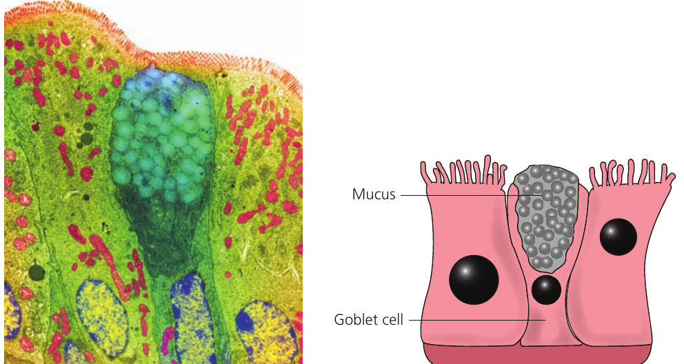
Goblet cells
Goblet cells are specialized secretory cells that produce the mucus lining the airways. Their name derives from their distinctive goblet shape — the wide upper portion (the "bowl") contains mucus ready for secretion, while the narrow base anchors the cell.
These cells are distributed between ciliated cells in the epithelium of the trachea, bronchi, and larger bronchioles. The mucus they secrete serves multiple functions:
- It traps particulate matter and microorganisms
- It maintains moisture in the airways
- It facilitates the movement of trapped particles by ciliary action
Smooth muscle and elastic fibres
Smooth muscle is found in the walls of the trachea, bronchi, and large bronchioles. It maintains baseline tone in the airways and can contract or relax to adjust airway diameter. During exercise, when oxygen demand increases, smooth muscle relaxation allows airway expansion and increased airflow.
Elastic fibres occur throughout all lung tissues, including the alveoli. These fibres provide the elasticity required for lung function. During inspiration, the lungs stretch as they fill with air. Expiration is predominantly passive — it relies on the elastic recoil of lung tissue as the stretched fibres return to their resting length.
This elastic property is essential for normal breathing mechanics. The elastic fibres enable passive expiration, meaning you don't need to actively force air out of your lungs during normal breathing.
Components of the gas exchange system
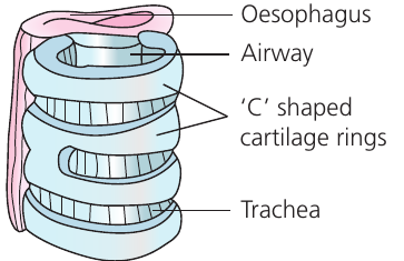
The trachea
The trachea is the widest tube in the respiratory system, serving as the main airway from the larynx to the bronchi. It does not participate in gas exchange; its sole function is to conduct air to the lungs.
The tracheal wall does not need to be especially thin (unlike gas exchange surfaces) nor particularly thick (as it plays no active role in ventilation). However, it must remain open at all times. Rather than developing a uniformly thick wall requiring substantial resources, the trachea has evolved C-shaped cartilage rings distributed at intervals along its length. These provide structural scaffolding while allowing flexibility in the softer tissue between rings.
The incomplete C-shape serves a specific purpose: the oesophagus runs directly posterior to the trachea and must expand when food passes through it. The gap in the cartilage ring faces the oesophagus, preventing friction between the oesophagus wall and rigid cartilage. The gap is bridged by tissue containing smooth muscle and elastic fibres.
The tracheal lining consists of ciliated epithelium containing goblet cells. Additionally, mucous glands lie beneath the epithelium. This abundant mucus production is particularly important in the trachea, as it is the first respiratory structure to encounter incoming air. Trapping most dust and bacteria at this point means they only need to travel the relatively short distance up the trachea to reach the oesophagus.
Bronchi
The bronchi branch from the trachea and share similar structural features, though with some modifications. They have smaller diameters and thinner walls than the trachea. The most significant structural difference is that bronchi possess complete rings of cartilage rather than C-shaped rings. This change is possible because the bronchi do not lie adjacent to the oesophagus, eliminating the need for an incomplete ring.
Like the trachea, bronchi have ciliated epithelium with goblet cells, mucous glands, smooth muscle, and elastic fibres.
Bronchioles
Bronchioles are smaller airways that branch extensively from the bronchi. They vary considerably in size and structure as they progressively divide and decrease in diameter toward the alveoli, ranging from approximately to in diameter.
The narrow diameter of bronchioles makes them more structurally self-supporting, eliminating the need for cartilage reinforcement. Consequently, no cartilage is present in bronchiolar walls.
Larger bronchioles retain goblet cells in their epithelium, but these are absent from smaller bronchioles. Similarly, cilia are progressively lost in smaller bronchioles. This reduction in mucociliary apparatus occurs because these smaller airways are deep within the lungs — inhaled air has already passed through multiple opportunities for particle trapping in more proximal airways.
Larger bronchioles contain smooth muscle and elastic fibres in their walls. The smooth muscle can contract or relax to adjust airway diameter, regulating airflow. Smaller bronchioles progressively lose smooth muscle, though elastic fibres persist even in the smallest bronchioles.
Alveoli
Alveoli are microscopic air sacs clustered at the terminal ends of the smallest bronchioles. These are the actual sites of gas exchange in the lungs.
Each alveolus has an extremely thin wall consisting of a single layer of epithelial cells. This minimal barrier facilitates rapid diffusion of oxygen and carbon dioxide. An extracellular matrix containing elastic fibres surrounds the epithelium, allowing alveoli to expand during inspiration and passively recoil during expiration.
Alveoli are extensively surrounded by capillary networks. These capillaries absorb oxygen that has diffused across the alveolar wall and deliver carbon dioxide for removal from the body. A thin film of watery fluid lines the alveolar interior, facilitating gas dissolution and diffusion while also creating surface tension.
Alveoli lack cartilage, smooth muscle, ciliated epithelium, goblet cells, and mucous glands. Their structure is optimized purely for gas exchange, with no role in air conduction or mucus clearance.
Comparison of airway structures
The following table summarizes the structural components present in different parts of the respiratory system:
| Structure | Cartilage | Smooth muscle | Elastic fibres | Ciliated epithelium | Goblet cells | Mucous glands |
|---|---|---|---|---|---|---|
| Trachea | ✓ | ✓ | ✓ | ✓ | ✓ | ✓ |
| Bronchi | ✓ | ✓ | ✓ | ✓ | ✓ | ✓ |
| Larger bronchioles | ✗ | ✓ | ✓ | ✓ | ✓ | ✗ |
| Smaller bronchioles | ✗ | ✓ | ✓ | ✗ | ✗ | ✗ |
| Smallest bronchioles | ✗ | ✗ | ✓ | ✗ | ✗ | ✗ |
| Alveoli | ✗ | ✗ | ✓ | ✗ | ✗ | ✗ |
Pattern Analysis:
This table reveals a clear pattern: structural complexity decreases as airways become smaller and approach the gas exchange surface. Cartilage disappears first (not needed in narrow, self-supporting tubes), followed by mucous glands, then goblet cells and cilia (as air is progressively cleaned), and finally smooth muscle (which has no role in the passive alveoli). Only elastic fibres persist throughout, reflecting their essential role in the mechanics of breathing.
Key Points to Remember:
- Cartilage provides structural support to prevent airway collapse in the trachea and bronchi; C-shaped rings in the trachea accommodate the adjacent oesophagus.
- Ciliated epithelium and goblet cells work together as a mucociliary escalator, with cilia beating to move mucus (containing trapped particles) upward toward the oesophagus.
- Elastic fibres are present throughout all airways and alveoli, enabling passive expiration through elastic recoil of stretched lung tissue.
- Structural complexity decreases progressively from trachea to alveoli: cartilage, mucous glands, goblet cells, cilia, and smooth muscle are sequentially lost, while elastic fibres remain present throughout.
- Alveoli are specialized purely for gas exchange, with single-layer epithelium, extensive capillary networks, and no conducting or cleaning structures.