Cellular Organelles (OCR A-Level Biology A): Revision Notes
Cellular Organelles
Eukaryotic cells contain numerous membrane-bound organelles that perform specialised functions. These organelles are suspended within the cytosol, a gel-like fluid that fills the cell. The cytoplasm refers to the entire region between the nucleus and the plasma membrane, including both the cytosol and the organelles it contains.
While often used interchangeably, cytosol and cytoplasm are distinct: the cytosol is just the fluid component, while the cytoplasm includes both the cytosol and all the organelles suspended within it.
The nucleus
The nucleus serves as the control centre of eukaryotic cells. It stores the cell's DNA, which acts as a template for RNA synthesis. Through RNA production, the nucleus indirectly regulates all cellular activities.
The nucleus is the defining feature of eukaryotic cells - it's what distinguishes them from prokaryotic cells, which lack a membrane-bound nucleus.
Structure
The nucleus is enclosed by a nuclear envelope (also called the nuclear membrane), which consists of two membranes. Each membrane has a similar structure to the plasma membrane. Nuclear pores are protein complexes that contain channels, allowing selective transport of molecules between the nucleus and cytoplasm.
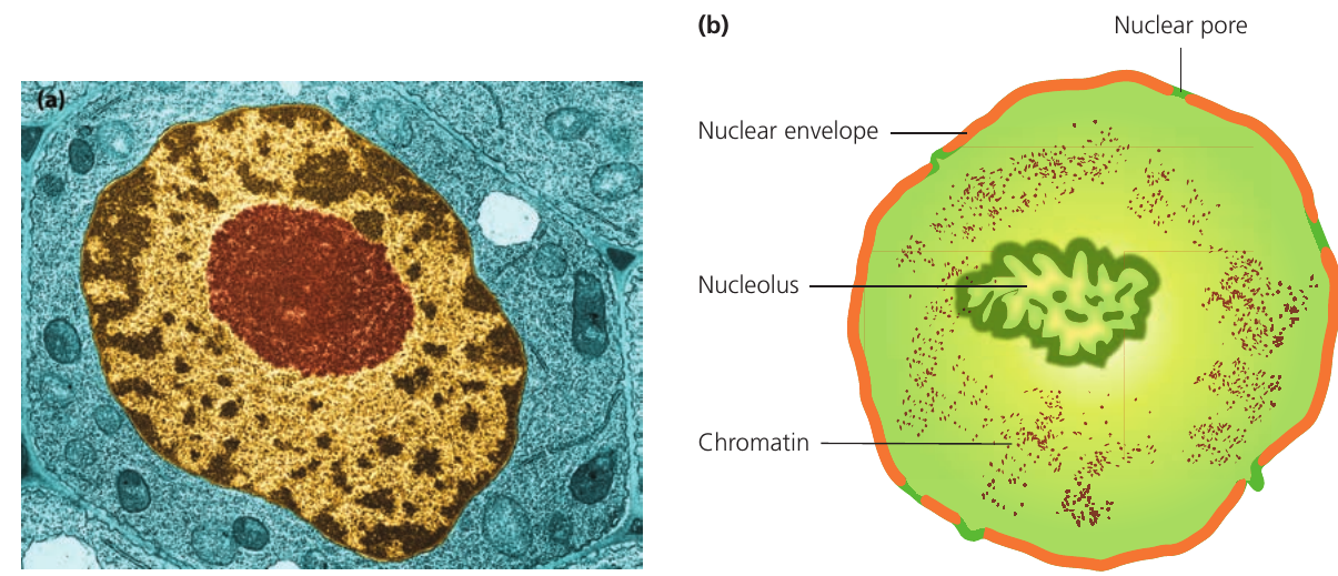
DNA within the nucleus exists in two forms depending on the cell's stage:
- Chromosomes: During cell division, DNA coils tightly and condenses into visible chromosomes
- Chromatin: At other times, DNA remains loosely distributed throughout the nucleus, appearing as grainy material under electron microscopy
The nucleolus appears as a darkly staining region within the nucleus. This specialised area constructs ribosomes, which are small enough to exit through nuclear pores and enter the cytoplasm.
The nucleolus is not surrounded by a membrane - it's simply a region where ribosomal RNA (rRNA) is synthesised and ribosomal subunits are assembled.
The endoplasmic reticulum
The endoplasmic reticulum forms part of the endomembrane system, which also includes the Golgi apparatus. This system of folded internal membranes creates channels throughout the cytoplasm, facilitating transport, processing and export of materials. The membranes connect with the outer nuclear membrane to form a continuous network.
The endoplasmic reticulum's extensive membrane network creates a vast surface area for cellular processes, similar to how the folded inner membrane of mitochondria increases surface area for respiration.
Rough endoplasmic reticulum
Rough endoplasmic reticulum (RER) gets its name from the ribosomes embedded in its membrane surface. These ribosomes synthesise proteins destined for secretion or transport outside the cell (proteins for internal use are made by free ribosomes in the cytoplasm).
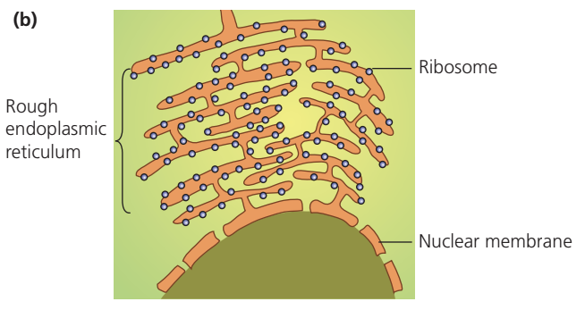
Newly synthesised proteins enter the lumen (internal space) of the RER, where they combine with carbohydrates. The modified proteins then travel to the Golgi apparatus for further processing. Cells that produce large quantities of proteins, such as enzyme-secreting or hormone-producing cells, contain extensive RER networks.
Protein destination matters: Ribosomes attached to the RER make proteins for export or insertion into membranes, while free-floating ribosomes in the cytoplasm make proteins for use within the cell itself.
Smooth endoplasmic reticulum
Smooth endoplasmic reticulum (SER) connects to the RER but lacks attached ribosomes. The SER specialises in lipid synthesis, including phospholipids for cell membranes. Cells that produce significant amounts of lipids, such as liver cells, contain abundant SER.
The SER also houses enzymes that detoxify lipid-soluble drugs and harmful metabolic products.
Liver cells are particularly rich in smooth ER because the liver is the body's main detoxification organ, constantly processing drugs and toxins to make them water-soluble for excretion.
Golgi apparatus
The Golgi apparatus (also called Golgi body or Golgi complex) consists of a series of flattened membranous sacs called cisternae. Unlike the endoplasmic reticulum, these cisternae are not interconnected. The structure divides into two functional sections: the cis Golgi network and the trans Golgi network.
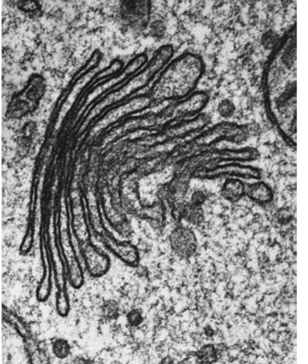
Function
The Golgi apparatus modifies proteins and lipids received from the RER, preparing them for secretion (the production and release of substances from a cell or gland). Vesicles pinched from the RER fuse with the cis face (innermost side) of the Golgi apparatus. After modification, materials move to the trans network, where new vesicles form. These vesicles transport their contents to the cell membrane, fuse with it, and release the materials outside the cell.
Additional functions include:
- Manufacturing lysosomes
- Synthesising cell wall materials in plant cells
Think of the Golgi apparatus as a cellular "post office" - it receives proteins, modifies and packages them, then addresses and ships them to their correct destinations.
Ribosomes
Ribosomes are protein-manufacturing organelles composed of ribosomal RNA (rRNA) and proteins. They exist in two separate components—a large subunit and a small subunit—which combine by attaching to messenger RNA (mRNA) when protein synthesis begins.
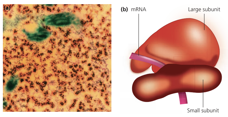
Distribution and types
Ribosomes occur in large numbers throughout cells, either:
- Free-floating in the cytoplasm (making proteins for internal cellular use)
- Attached to RER membranes (making proteins for secretion)
Eukaryotic cells contain 80S ribosomes, which are larger than the 70S ribosomes found in prokaryotic cells. The 'S' refers to Svedberg units, which measure sedimentation rate during centrifugation—higher values indicate larger, faster-sedimenting particles.
Don't confuse ribosome size with cell type: 80S ribosomes are found in eukaryotic cells, while 70S ribosomes are characteristic of prokaryotic cells. This distinction becomes important when studying mitochondria and chloroplasts, which contain 70S ribosomes despite being in eukaryotic cells.
Mitochondria
Mitochondria are double-membrane organelles responsible for aerobic respiration. Cells with high energy demands, such as muscle cells and nerve cells, contain particularly large numbers of mitochondria.
Structure
The inner membrane folds inward to form cristae (singular: crista), creating a large surface area for respiratory enzymes. The space enclosed by the inner membrane is called the matrix.
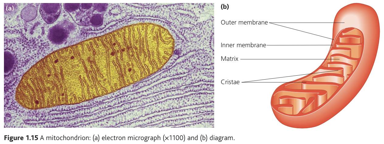
The extensive folding of the inner membrane into cristae is an excellent example of the relationship between structure and function - the large surface area allows more respiratory enzymes to be embedded, increasing the rate of ATP production.
Evolutionary significance
Mitochondria contain their own DNA and 70S ribosomes (similar to prokaryotic ribosomes). This similarity to bacteria provides evidence for the endosymbiotic theory—the idea that mitochondria evolved from ancient prokaryotic organisms.
The presence of 70S ribosomes and circular DNA in mitochondria strongly supports the endosymbiotic theory, suggesting these organelles were once free-living prokaryotes that were engulfed by early eukaryotic cells and developed a mutually beneficial relationship.
Chloroplasts
Chloroplasts are membrane-bound organelles found exclusively in plant cells. They absorb light energy to drive photosynthesis.
Structure
A double outer membrane surrounds the chloroplast. Inside, a complex arrangement of internal membranes forms thylakoids—flattened sac-like structures that group into stacks called grana (singular: granum). Intergranal lamellae connect these stacks. Chlorophyll pigments, which capture light energy, are embedded in the thylakoid membranes.
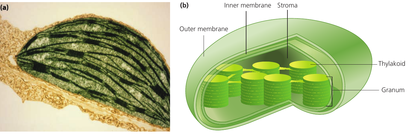
The space between grana is filled with stroma, a thick fluid. Different stages of photosynthesis occur in different locations: some in the grana, others in the stroma.
The light-dependent reactions of photosynthesis occur in the thylakoid membranes where chlorophyll is located, while the light-independent reactions (Calvin cycle) take place in the stroma.
Like mitochondria, chloroplasts contain their own DNA and 70S ribosomes, suggesting they too may have evolved from prokaryotic organisms.
Remember: Chloroplasts are found ONLY in plant cells and some protists, never in animal cells. This is a common exam point!
Lysosomes
Lysosomes specialise in digesting cellular material. Each lysosome contains approximately ``50 different hydrolytic enzymes capable of breaking down proteins, nucleic acids, carbohydrates and lipids.
Function and safety
A single membrane separates lysosomal enzymes from the rest of the cytoplasm. The enzymes function optimally in the acidic environment inside lysosomes. If enzymes escape into the neutral cytoplasm, they become relatively ineffective, protecting the cell from damage.
The acidic environment inside lysosomes (pH ~5) is a crucial safety mechanism. If the lysosome membrane breaks and enzymes leak into the neutral cytoplasm (pH ~7), they become much less active, preventing them from digesting the entire cell.
Lysosomes digest material from various sources:
- External materials (e.g. bacteria)
- Internal materials (e.g. damaged or aged organelles)
Under electron microscopy, lysosomes appear as darkly stained structures without clearly distinguishable internal features.
Plasma membrane
The plasma membrane forms the cell's outer boundary. It acts as a selective barrier, controlling the movement of substances into and out of the cell. This control arises from:
- Differential permeability to different biological molecules
- Protein carriers that actively pump substances according to cellular needs
The plasma membrane's selective permeability is essential for maintaining cellular homeostasis - it allows nutrients in and waste products out while keeping the cell's internal environment stable.
Other cellular membranes (around organelles, forming the endoplasmic reticulum, surrounding vacuoles) share the basic structure of phospholipids and proteins, though specific protein compositions vary.
Centrioles and microtubules
Centrioles are cylindrical structures composed of microtubules (small tubular structures made of the protein tubulin arranged in spirals). Each centriole contains nine triplets of microtubules arranged in a ring pattern.
Centrioles occur in pairs near the nucleus and are found only in animal cells. While once thought to organise spindle formation during cell division, we now know that centrosomes perform this role. The spindle is a system of microtubules that separates chromosomes during cell division.
Plant cells lack centrioles but can still form spindle fibres during cell division. The centrosomes, not the centrioles themselves, are the key organising centres for spindle formation.
Flagella and cilia
Flagella (singular: flagellum) and cilia (singular: cilium) are surface structures composed of microtubules. They create currents through their movement, either propelling the cell itself or moving liquids across the cell surface.
Distribution and function
- Flagella: Found singly or in pairs in some single-celled organisms and form the 'tail' of sperm cells. They propel cells through their environment
- Cilia: Always present in large numbers on both single-celled organisms and cells of multicellular animals. They move liquids across cell surfaces
Structure
Both flagella and cilia share the same internal structure: nine pairs of microtubules arranged in a ring surrounding two central microtubules. The cell membrane extends over these structures. The only differences between flagella and cilia are length (flagella are longer) and number (cilia occur in greater numbers).
The "9+2" arrangement of microtubules (nine pairs around the outside, two in the centre) is characteristic of eukaryotic flagella and cilia. This structure is remarkably consistent across different organisms.
Prokaryotic flagella differ significantly—they are smaller with a simpler structure than eukaryotic flagella.
Don't confuse prokaryotic and eukaryotic flagella! Despite sharing the same name, prokaryotic flagella are completely different structures - they're made of a protein called flagellin and rotate like a propeller, while eukaryotic flagella contain microtubules and beat in a wave-like motion.
Cell wall
The cell wall is an extracellular layer that provides structural support and prevents cells from bursting when water enters by osmosis.
Composition varies by organism type
In eukaryotes:
- Plants and algae: Cellulose (chains of glucose molecules)
- Fungi: Chitin (similar to cellulose but containing nitrogen)
In prokaryotes:
- Bacteria: Murein (a polymer of sugars and amino acids)
The cell wall prevents cells from bursting in hypotonic solutions, but it doesn't stop water entering - osmosis still occurs. The wall simply provides mechanical strength to resist the pressure that builds up inside the cell.
Cell fractionation
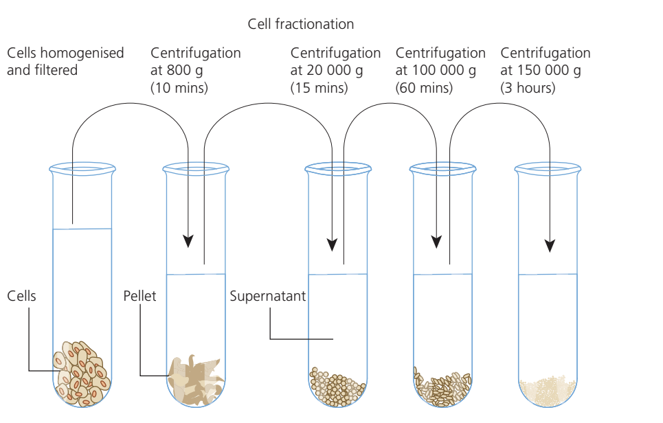
Cell fractionation is a laboratory technique for isolating cellular organelles. The process involves:
- Homogenisation: Breaking up cells in an ice-cold isotonic solution (same solute concentration as cell contents)
- Filtration: Removing large debris
- Centrifugation: Spinning the homogenate at progressively higher speeds
Principle
Organelles sediment at different rates depending on their mass. Heavier organelles compact into pellets at lower centrifugal speeds, while lighter organelles require higher speeds. By increasing the centrifugal force stepwise, different organelles can be isolated in sequence.
Typical Fractionation Sequence:
Step 1: Low speed centrifugation (~1,000 g)
- Nuclei pellet first (heaviest organelles)
- Supernatant contains remaining organelles
Step 2: Medium speed centrifugation (~10,000 g)
- Mitochondria and chloroplasts pellet
- Supernatant still contains lighter organelles
Step 3: High speed centrifugation (~100,000 g)
- Ribosomes and membrane fragments pellet
- Final supernatant contains only soluble proteins
The ice-cold temperature prevents enzyme activity that could damage organelles. The isotonic solution prevents osmotic damage to organelles.
Both temperature and solution concentration are critical for successful cell fractionation:
- Cold temperature: Reduces enzyme activity that would digest organelles
- Isotonic solution: Prevents organelles from bursting (hypotonic) or shriveling (hypertonic) due to osmosis
Approximate organelle sizes
| Organelle | Approximate size () |
|---|---|
| Nucleus (diameter) | |
| Ribosome (diameter) | |
| Mitochondrion (length diameter) | |
| Chloroplast (length width) |
Key Points to Remember:
- The nucleus contains DNA and controls cellular activities through RNA production; it is surrounded by a double nuclear envelope with nuclear pores
- Rough ER has attached ribosomes and synthesises proteins for secretion, while smooth ER lacks ribosomes and produces lipids
- The Golgi apparatus modifies and packages proteins and lipids from the ER into secretory vesicles
- Mitochondria perform aerobic respiration and contain their own DNA and 70S ribosomes, suggesting prokaryotic origins
- Chloroplasts (plant cells only) carry out photosynthesis; their thylakoid membranes contain chlorophyll and stack into grana
- Lysosomes contain hydrolytic enzymes that digest cellular material in acidic conditions
- Both mitochondria and chloroplasts contain 70S ribosomes and their own DNA, providing evidence for endosymbiotic evolution from prokaryotes
- Cell fractionation separates organelles by mass using centrifugation at progressively higher speeds in ice-cold, isotonic conditions