Organelles Working Together (OCR A-Level Biology A): Revision Notes
Organelles Working Together
Division of labour in cells
Cells contain multiple organelles, each performing specialized functions. Many cellular processes require several organelles to work in a coordinated sequence. A key example is the production and secretion of proteins, which involves six different cellular components working together.
The concept of division of labour means that each organelle has a specific role to perform, and these roles must be coordinated for the cell to function effectively. This is similar to how workers in a factory each have specialized tasks that contribute to the final product.
Protein synthesis and secretion pathway
Cells that produce proteins for secretion (such as enzymes or hormones) must coordinate the activity of multiple organelles. The process follows a specific sequence involving the nucleolus, nucleus, rough endoplasmic reticulum, Golgi body, vesicles, and plasma membrane.
Worked Example: The Six-Step Protein Synthesis and Secretion Pathway
This pathway demonstrates how organelles work as an integrated system to produce and secrete proteins:
Step 1: Ribosome production The nucleolus produces ribosomes, which are essential for protein synthesis. These ribosomes will be used on the rough endoplasmic reticulum.
Step 2: mRNA synthesis The nucleus synthesizes messenger RNA (mRNA), which carries the genetic instructions needed by ribosomes to assemble proteins.
Step 3: Protein assembly Ribosomes attached to the rough endoplasmic reticulum (RER) use the mRNA template to synthesize proteins. The RER provides the site where proteins destined for secretion are made.
Step 4: Initial protein processing The RER processes the newly synthesized proteins, modifying them and preparing them for transport. These proteins are packaged into transport vesicles that bud off from the RER.
Step 5: Further processing and packaging Transport vesicles carry the proteins to the Golgi body (also called Golgi apparatus). The Golgi body further processes and modifies the proteins, then packages them into secretory vesicles.
Step 6: Secretion Secretory vesicles move to the plasma membrane. The vesicles fuse with the plasma membrane, releasing their protein contents outside the cell through a process called exocytosis.
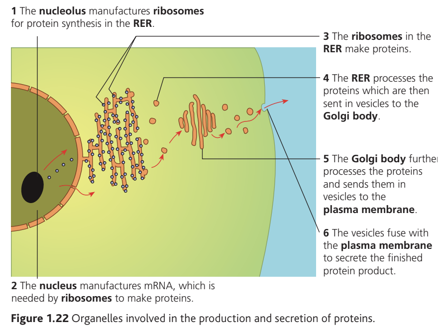
This pathway demonstrates how organelles work as an integrated system. Each organelle completes a specific task, and the product passes to the next organelle in the sequence, much like a production line in manufacturing.
The cytoskeleton
The cytoskeleton is a network of protein filaments that extends throughout the cytoplasm. It consists of two main types of structure: microtubules and microfilaments.
Structure and composition
Microtubules are hollow, tubular structures made primarily from the protein tubulin. Their tubular design provides rigidity and forms channels for transport.
Microfilaments are solid strands composed mainly of the protein actin. These thinner filaments provide flexibility and contractile properties.
Remember the structural differences and protein composition:
- Microtubules = tubular (hollow) structure made of tubulin protein
- Microfilaments = solid strands made of actin protein
Memory aid: "TUBULIN in TUBules" and "ACTIN in filAments"
Functions of the cytoskeleton
The cytoskeleton performs three essential roles:
Cellular movement Microtubules form the internal structure of cilia and flagella, hair-like projections that extend from the cell surface. Movement of these structures allows cells to swim through fluids, or to move fluids across the cell surface (such as mucus movement in airways).
Intracellular movement The cytoskeleton creates transport pathways within the cell. Intracellular (meaning within a cell) movement occurs when organelles and vesicles travel along these protein tracks from one location to another. During cell division, microtubules form the spindle apparatus that moves chromosomes to opposite ends of the dividing cell.
Intracellular means "within a cell" – this distinguishes movement happening inside the cell from movement of the whole cell through its environment.
Strengthening and support The cytoskeleton acts as an internal scaffolding system. It supports organelles in their correct positions and maintains the cell's shape, preventing the cell from collapsing under external pressure.
Prokaryotic and eukaryotic cells
All cells fall into one of two fundamental categories: prokaryotic or eukaryotic.
Prokaryotic cells include all bacteria and blue-green bacteria (cyanobacteria). These are the simplest cell type.
Eukaryotic cells include all other organisms: animals, plants, fungi, and protists. These cells are more complex and contain specialized compartments.
Key structural differences
The table below compares the main structural features:
| Feature | Eukaryotic cell | Prokaryotic cell |
|---|---|---|
| Nucleus | Present – contains DNA within a membrane-bound compartment | Absent – no nuclear membrane |
| DNA organization | Linear chromosomes located in the nucleus | Circular DNA molecule free in the cytoplasm |
| Cell wall composition | When present: cellulose (plants) or chitin (fungi) | Made of murein (peptidoglycan) |
| Membrane-bound organelles | Present – includes mitochondria, ER, Golgi apparatus | Absent – no compartmentalization |
| Ribosome size | Large 80S ribosomes | Small 70S ribosomes |
| Capsule | Absent | Present – protective outer layer |
| Pili | Absent | Sometimes present – used for attachment or DNA transfer |
| Cell size | Larger: – | Smaller: less than |
Critical Difference – Ribosome Size:
- Eukaryotic cells contain 80S ribosomes (larger)
- Prokaryotic cells contain 70S ribosomes (smaller)
This difference is important because certain antibiotics specifically target 70S ribosomes, allowing them to kill bacteria without harming human cells.
Prokaryotic cell structure
Bacterial cells contain several distinctive features. The cell wall made of murein provides structural support and protection. Many bacteria possess a capsule, an additional protective layer outside the cell wall that helps the bacterium evade immune responses.
Plasmids are small, circular DNA molecules separate from the main circular chromosome. They often carry genes for antibiotic resistance and can be transferred between bacteria.
Pili (singular: pilus) are hair-like protein projections used for attachment to surfaces or for transferring DNA between cells during conjugation.
Flagella (singular: flagellum) are longer whip-like structures that rotate to propel the bacterium through liquid environments.
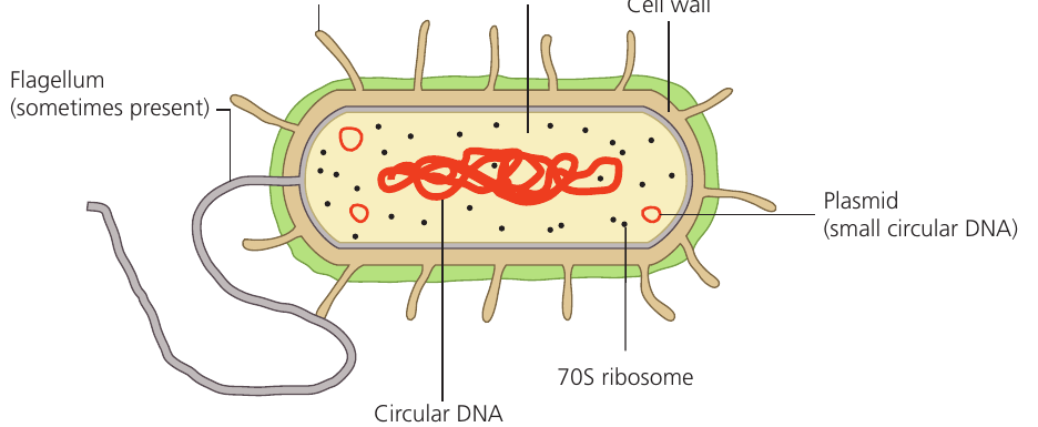
The cytoplasm contains 70S ribosomes that synthesize proteins. The main circular DNA molecule is not enclosed within a nucleus but exists freely in a region called the nucleoid.
Protoplast refers to the part of a cell (in bacteria or plants) that is inside the cell wall. In bacteria, this includes the plasma membrane and all internal contents.
Plant and animal cells
Both plant and animal cells are eukaryotic, but they show important structural differences.
Plant cell features
Plant cells possess several structures not found in animal cells:
Cellulose cell wall A rigid wall made of cellulose fibres surrounds the plasma membrane. This provides structural support, allows high internal pressure (turgor), and prevents the cell from bursting.
Plasmodesmata These are narrow channels that penetrate through the cell wall. Plasmodesmata (singular: plasmodesma) allow cytoplasm to connect between adjacent plant cells, enabling direct communication and transport of materials. The protoplast (the part of the cell inside the cell wall) of one cell thus connects to neighboring protoplasts.
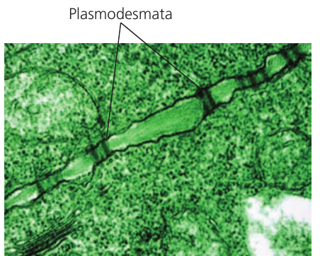
Plasmodesmata create a continuous network of cytoplasm throughout plant tissues, allowing cells to share resources and coordinate their activities. This is fundamentally different from animal tissues where cells are completely separate from one another.
Chloroplasts These organelles carry out photosynthesis, converting light energy into chemical energy. Only cells in photosynthetic tissues (leaves, stems) contain chloroplasts; root cells and other non-photosynthetic plant cells lack them.
Large central vacuole A membrane-bound sac filled with cell sap (water and dissolved substances) occupies most of the plant cell volume. The vacuole maintains turgor pressure, stores nutrients, and breaks down waste products.
Animal cell features
Animal cells contain two structures rarely or never found in plant cells:
Centrioles These cylindrical structures made of microtubules play a role in organizing the spindle during cell division. Plant cells can undergo cell division without centrioles.
Flagella Some animal cells (such as sperm cells) possess flagella for movement. Flagella are extremely rare in plant cells, occurring only in one primitive group (cycads).
Key Points to Remember:
-
Organelles cooperate in sequences – complex processes like protein secretion require multiple organelles working in a specific order: nucleolus → nucleus → RER → Golgi body → vesicles → plasma membrane.
-
The cytoskeleton has three functions – cellular movement (cilia/flagella), intracellular transport (organelles and vesicles along protein tracks), and structural support (maintaining cell shape).
-
Prokaryotic cells lack membrane-bound organelles – bacteria have no nucleus, mitochondria, or endoplasmic reticulum, but possess unique features: circular DNA, murein cell wall, 70S ribosomes, and sometimes capsules, pili, or plasmids.
-
Microtubules are tubular, microfilaments are solid – microtubules contain tubulin protein and form hollow tubes; microfilaments contain actin protein and form solid strands.
-
Plant cells have distinctive features – cellulose cell wall with plasmodesmata, chloroplasts (in photosynthetic cells), and a large central vacuole distinguish plant cells from animal cells.