Proteins and Nucleic Acids (OCR A-Level Biology A): Revision Notes
Proteins
Introduction
Proteins are large biological molecules with diverse and vital roles in living organisms. They account for approximately of the organic molecules in cells (excluding water).
Elemental composition: Proteins contain carbon (C), hydrogen (H), oxygen (O) and nitrogen (N). Some proteins also contain sulfur (S).
Remember these five elements with the mnemonic CHONS!
Functions of proteins:
- Structural: Components of cytoplasm, muscle fibres, collagen in skin and bone, keratin in hair, elastin in connective tissue
- Catalytic: All enzymes are proteins
- Transport: Carrier molecules in cell membranes, haemoglobin for oxygen transport
- Communication: Many hormones (e.g. insulin)
- Defence: Antibodies in the immune system
- Growth and repair: Building blocks for new cells and tissues
Amino acids: the building blocks
Proteins are polymers composed of smaller subunits called amino acids. These amino acids act as monomers that link together to form protein chains.
General structure of amino acids
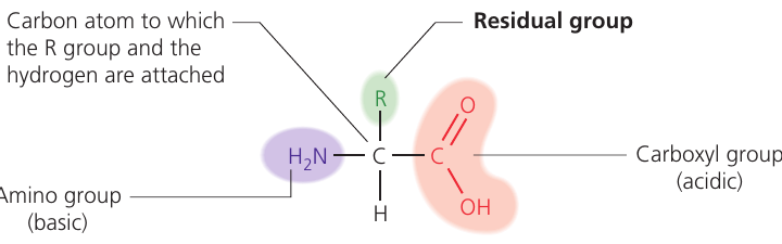
All amino acids share a common basic structure containing four groups attached to a central carbon atom:
- Amino group (–NH₂): Basic functional group (shown in purple in diagrams)
- Carboxyl group (–COOH): Acidic functional group (shown in pink/red in diagrams)
- Hydrogen atom (H)
- R group (residual group): Variable side chain (shown in green in diagrams)
The R group determines amino acid identity
There are 20 different amino acids that make up proteins in living organisms. The R group is the only part that differs between amino acids, giving each its unique properties.
The R group is critical! It determines:
- The identity of each amino acid
- How amino acids interact with each other
- The properties of the final protein structure
R group characteristics:
- Size: Ranges from a single hydrogen atom (glycine) to large complex groups
- Polarity: Can be polar (hydrophilic) or non-polar (hydrophobic)
- Charge: May be acidic, basic, or neutral
Examples of amino acids:
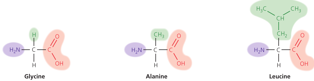
- Glycine: Smallest amino acid with H as its R group
- Alanine: Contains a methyl group (–CH₃) as its R group
- Leucine: Has a larger branched hydrocarbon R group
Types of R groups
Polar/charged R groups (hydrophilic):
- Acidic polar amino acids: e.g. aspartic acid with –CH₂–COOH
- Basic polar amino acids: e.g. arginine with (–CH₂)₃–HN–CNH–NH₂
- Polar uncharged amino acids: e.g. serine with –CH₂OH
Non-polar R groups (hydrophobic):
- e.g. valine with –CH(CH₃)₂
The chemical properties of R groups determine how amino acids interact with each other and influence the final protein structure. Hydrophilic R groups tend to be on the protein surface (interacting with water), while hydrophobic R groups tend to be buried inside the protein structure.
Peptide bond formation
Condensation reaction
Two amino acids join together through a condensation reaction (also called dehydration synthesis):
- The hydroxyl group (–OH) from the carboxyl group of one amino acid combines with a hydrogen atom (H) from the amino group of another
- One molecule of water (H₂O) is released
- A covalent bond called a peptide bond forms between the carbon of one amino acid and the nitrogen of the next
The resulting molecule is called a dipeptide.
Hydrolysis reaction
The peptide bond can be broken by hydrolysis:
- One molecule of water (H₂O) is added
- The peptide bond breaks
- Two separate amino acids are reformed
These reactions are reversible:
- Condensation builds peptide bonds (removes water)
- Hydrolysis breaks peptide bonds (adds water)
This is fundamental to both protein synthesis and protein digestion!
Polypeptides
A polypeptide is a chain of many amino acids joined by peptide bonds.
Key features:
- Each polypeptide has an N-terminal (amino end) and a C-terminal (carboxyl end)
- Polypeptides vary in:
- Number of amino acids (from tens to hundreds)
- Types of amino acids present
- Sequence (order) of amino acids
Once amino acids are incorporated into a polypeptide, they are often referred to as amino acid residues.
The specific sequence, number and type of amino acids determine the properties and function of the resulting protein.
Levels of protein structure
Protein molecules are organized at different structural levels. Understanding these levels is essential for understanding how proteins function.
Mnemonic: "Please Stay Totally Quiet"
- Primary
- Secondary
- Tertiary
- Quaternary
Primary structure
The primary structure is the specific sequence of amino acids in the polypeptide chain, including:
- The exact order of amino acids
- The number of amino acids
- The position of any disulfide bonds
The primary structure is determined by genetic information (DNA sequence) and is unique to each protein. Even a single change in one amino acid can alter or completely disrupt protein function.
This is why genetic mutations that change amino acid sequences can have such severe consequences!
Secondary structure
The secondary structure forms when the polypeptide chain folds or coils due to hydrogen bonding between amino acids at different positions along the chain.
Two main types of secondary structure exist:
Alpha helix (α-helix):
- The polypeptide chain coils into a right-handed spiral (like a spring)
- Stabilized by hydrogen bonds between the C=O of one amino acid and the N–H of an amino acid further along the chain
- Creates a rod-like structure
Beta pleated sheet (β-sheet):
- The polypeptide chain folds back and forth like a concertina
- Adjacent parallel or antiparallel strands are held together by hydrogen bonds
- Creates a sheet-like structure with a pleated appearance
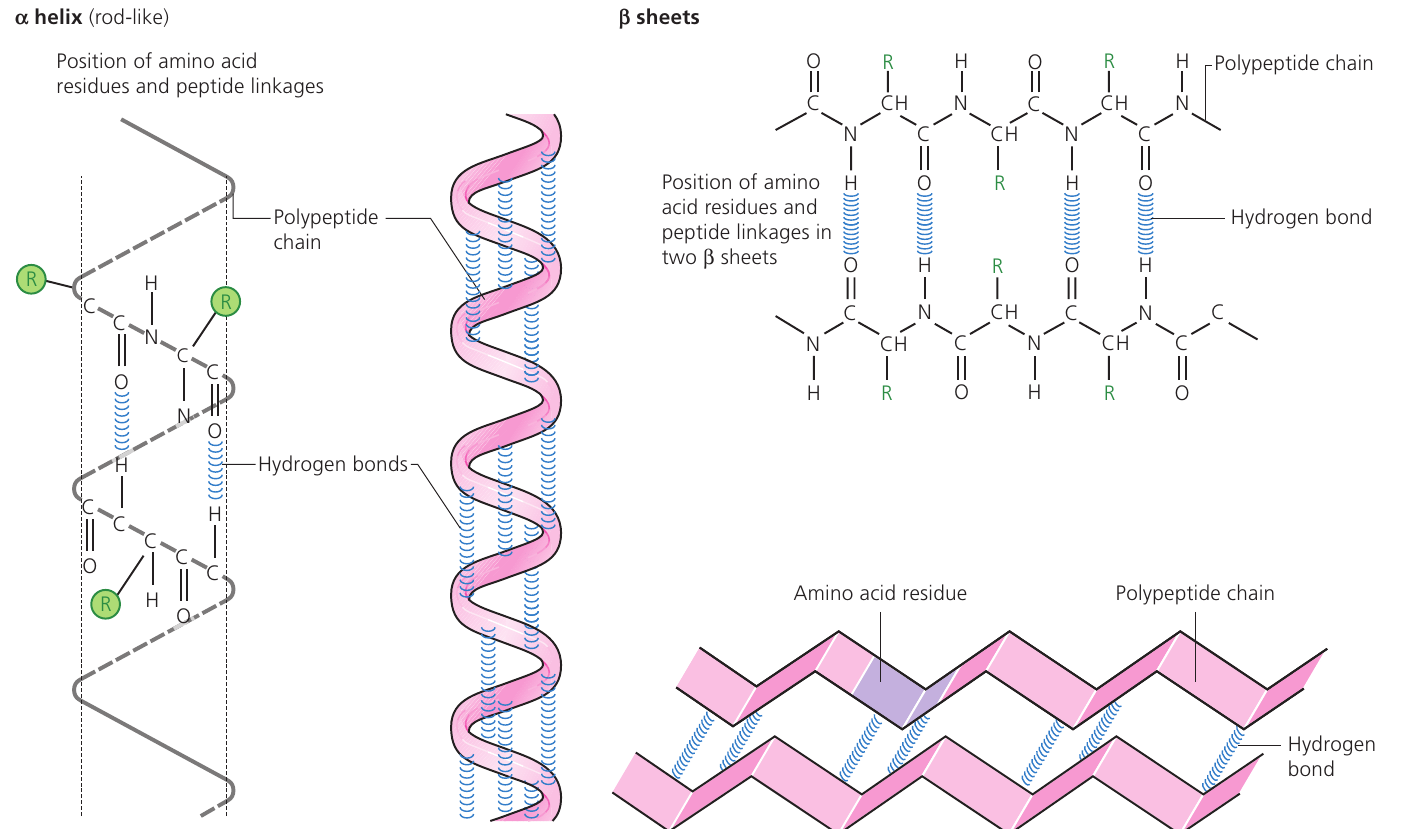
Hydrogen bonds are individually weak, but their large numbers provide significant stability to the protein structure.
Tertiary structure
The tertiary structure is the complex three-dimensional shape that forms when the secondary structure folds and coils further.
This 3D shape is critical for protein function and is maintained by several types of bonds and interactions:
Types of bonds in tertiary structure:
- Hydrogen bonds: Weak attractions between slightly charged groups (δ⁺ and δ⁻) on different amino acids
- Ionic bonds: Attractions between oppositely charged R groups (positive and negative)
- Hydrophobic interactions: Association of non-polar R groups that exclude water
- Disulfide bonds: Strong covalent bonds between sulfur atoms in cysteine residues (–S–S–)
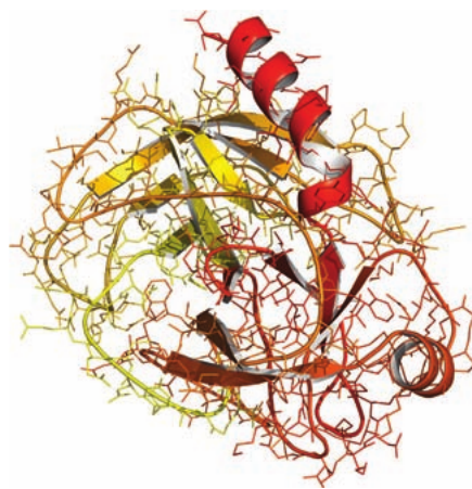
The 3D shape determines function!
If the tertiary structure is disrupted (by heat, pH changes, or chemicals), the protein becomes denatured and loses its function. This is why maintaining correct conditions is crucial for protein activity.
Two main tertiary structure types:
Globular proteins:
- Fold into compact, roughly spherical shapes
- Generally soluble in water
- Examples: enzymes, antibodies, hormones (insulin), haemoglobin
- Function: metabolic roles requiring 3D shape (e.g. enzyme active sites)
Fibrous proteins:
- Form long, rope-like or strand-like structures
- Generally insoluble in water
- Examples: collagen, keratin, elastin, fibrin
- Function: structural support and strength
Quaternary structure
Some proteins consist of more than one polypeptide chain. The quaternary structure describes how these multiple chains (subunits) associate to form a functional protein.
Key features:
- Two or more polypeptide chains held together by hydrogen bonds, ionic bonds, and disulfide bonds
- The protein only functions when all subunits are correctly assembled
- Subunits may be identical or different
- Some quaternary proteins include non-protein components (prosthetic groups)
Not all proteins have quaternary structure—only those composed of multiple polypeptide chains. Proteins with a single polypeptide chain (like many enzymes) only have primary, secondary, and tertiary structures.
Examples of proteins
Globular protein: haemoglobin
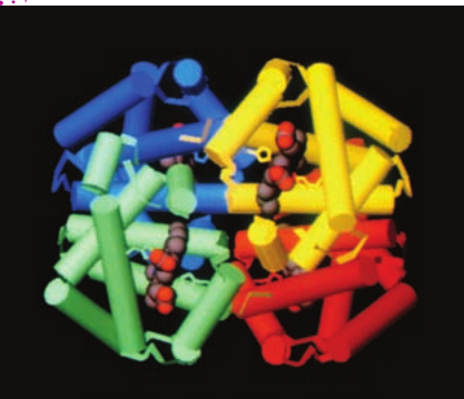
Structure:
- Contains four polypeptide chains: two α-chains and two β-chains
- Each chain associates with a haem group (prosthetic group)
- The haem group contains an iron ion (Fe²⁺ or Fe³⁺) within a porphyrin ring
- The four chains are held together by hydrogen bonds, ionic bonds, and hydrophobic interactions
- Forms a compact, roughly spherical (globular) shape
Classification: Haemoglobin is a conjugated protein because it contains a non-protein prosthetic group (haem).
The term prosthetic group refers to any non-protein component that is essential for a protein's function. The haem group is also called a cofactor in enzyme terminology.
Function: Oxygen transport in red blood cells. Each iron ion can bind one oxygen molecule, allowing each haemoglobin molecule to carry four oxygen molecules.
Solubility: Soluble in water/blood plasma
Fibrous protein: collagen

Structure:
- Contains three polypeptide chains twisted around each other like a rope or plait
- Each chain consists of a repeating sequence of three amino acids
- One of these amino acids is always glycine (the smallest amino acid)
- The chains are held together by numerous hydrogen bonds and covalent bonds
- Multiple collagen molecules associate to form collagen fibrils, which combine to form collagen fibres
Helical structure: Left-handed helix (unlike the right-handed α-helix)
The presence of glycine at every third position is crucial because its small size allows the chains to pack tightly together in the triple helix structure.
Function: Provides strength and structural support in:
- Artery walls (withstands blood pressure)
- Tendons (connects muscle to bone)
- Bone and cartilage (structural framework)
- Skin (provides elasticity and strength)
Solubility: Insoluble in water
Comparison of haemoglobin and collagen
| Feature | Haemoglobin | Collagen |
|---|---|---|
| Protein type | Globular | Fibrous |
| Number of polypeptides | 4 (two α, two β chains) | 3 |
| 3D structure | Compact, ball-like shape | Long, twisted fibrous structure |
| Helical structure | Right-handed α-helix | Left-handed helix |
| Solubility | Soluble in water | Insoluble in water |
| Amino acid variety | Contains most of the 20 amino acids | Limited variety; high glycine content with repeating triplet sequence |
| Prosthetic group | Contains haem group with iron | No prosthetic group |
| Biological role | Oxygen transport | Structural support in arteries, tendons, cartilage, bone, and skin elasticity |
Other important proteins
Insulin:
- Globular protein and hormone
- Composed of two polypeptide chains: chain A (21 amino acids) and chain B (30 amino acids)
- Chains held together by three disulfide bridges
- First protein to have its amino acid sequence determined (Fred Sanger, 1955)
- Function: regulates blood glucose levels
Enzymes:
- All enzymes are globular proteins
- Examples: amylase, catalase, trypsin
- Require specific 3D shape to form active sites
- Active site is complementary to specific substrate molecules
- Function: catalyse (speed up) biochemical reactions
The active site of an enzyme is formed by the tertiary structure. If the shape changes (denaturation), the enzyme loses its catalytic function. This is why temperature and pH are so critical for enzyme activity!
Keratin:
- Fibrous protein found in hair and nails
- Provides structural strength
Elastin:
- Fibrous protein in connective tissue, skin, tendons, and bone
- Can stretch and return to original shape
- Provides elasticity
Testing for proteins: the biuret test
The biuret test is a qualitative biochemical test that detects the presence of proteins.
Principle: Copper ions (Cu²⁺) in alkaline solution react with peptide bonds to form a coloured complex.
Procedure:
- Add equal volumes of protein solution and sodium hydroxide (NaOH)
- Shake to mix thoroughly
- Add a small amount of copper sulfate solution drop by drop
- Shake between each addition
- Observe colour change
Results:
| Test solution | Colour before reagents | Colour after reagents | Interpretation |
|---|---|---|---|
| Protein present | Cloudy suspension | Mauve/purple | Positive result |
| No protein (control) | Cloudy suspension | Blue (copper sulfate colour) | Negative result |
Key Points about the Biuret Test:
- The test is qualitative (indicates presence or absence) rather than quantitative (measures concentration)
- To make it quantitative, a colorimeter would be needed to measure the intensity of the mauve colour against known standards
- The mauve/purple colour indicates the presence of peptide bonds (and therefore proteins or polypeptides)
- The more intense the colour, the more peptide bonds present
Key Points to Remember:
- Amino acids are the monomers of proteins; 20 different types exist, varying in their R groups
- Peptide bonds form between amino acids through condensation reactions (releasing water) and can be broken by hydrolysis (adding water)
- Proteins have up to four levels of structure: primary (sequence), secondary (α-helix or β-sheet), tertiary (3D shape), and quaternary (multiple chains)
- The 3D shape of a protein determines its function—shape changes can disrupt or destroy function
- Globular proteins (e.g. enzymes, haemoglobin) are compact and soluble, while fibrous proteins (e.g. collagen) are long, rope-like, and provide structural support
- Use the mnemonic "Please Stay Totally Quiet" to remember the four levels of protein structure
- The biuret test produces a mauve/purple colour in the presence of proteins