Structure of the Heart (Leaving Cert Biology): Revision Notes
📚 Revision Notes
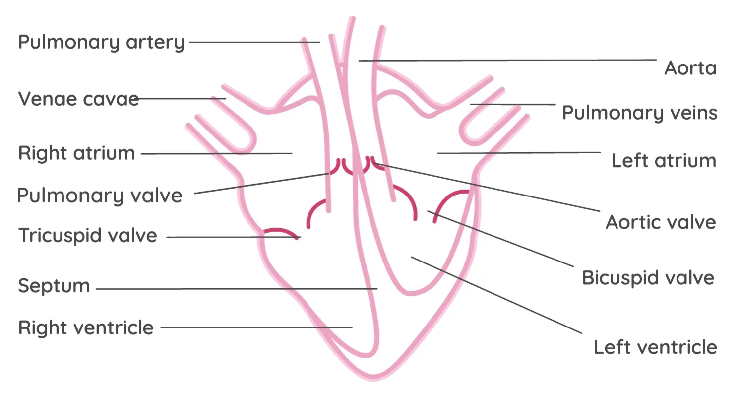
Structure of the Heart
- The heart is located between the lungs and above the diaphragm.
- The heart is made of cardiac muscle, which never tyres. It is under involuntary control.
- It is surrounded by a membrane called the pericardium. This reduces friction between the heart and nearby organs.
- Chambers:
- Two upper chambers: Atria
- Two lower chambers: Ventricles
- Ventricles:
- The right ventricle pumps blood to the lungs.
- The left ventricle is thicker because it pumps blood all around the body.
- The heart is separated into left and right sides by a wall called the septum.
- Valves:
- The atria and ventricles are separated by valves, which prevent the backflow of blood.
- The tricuspid valve is between the right atrium and right ventricle.
- The bicuspid valve is between the left atrium and left ventricle.
- There are two semilunar valves.
- One where the aorta leaves the left ventricle and another where the pulmonary artery leaves the right ventricle.
- Key Blood Vessels:
- Aorta: Largest artery, carries oxygenated blood from the left ventricle to the body.
- Vena cava: Largest vein, carries deoxygenated blood from the body to the heart.
- Pulmonary artery: Carries deoxygenated blood from the right ventricle to the lungs.
- Pulmonary vein: Brings oxygenated blood from the lungs to the heart.
TIP: When drawing a diagram of the heart, remember that the left and right sides are reversed. Label the left side of the diagram as "Right" and the right side as "Left".
infoNote
Note:
- Your heart is roughly the size of your fist.
- It is positioned slightly to the left of the midline in the chest, and the left lung is slightly smaller than the right lung to accommodate the space taken up by the heart.
infoNote
Cardiac muscle is unique because it does not tyre.
infoNote
L O R D
L O : Left side carries Oxygenated blood.
R D: Right side carries Deoxygenated blood.