The Heart (Leaving Cert Biology): Revision Notes
The Heart
Introduction to the heart
The heart is a remarkable muscular organ that works as the body's central pump. Located between the two lungs in the chest cavity, it sits slightly to the left-hand side and is about the size of a clenched fist. This vital organ works tirelessly, contracting approximately 100,000 times each day and pumping between 5 and 20 litres of blood every minute to keep your body functioning properly.
The heart never gets a break - it beats continuously from before you're born until the day you die, making it one of the most hardworking organs in your body!
Structure of the heart
The heart is constructed from a special type of muscle called cardiac muscle. This muscle tissue is unique because it can contract rhythmically without getting tired easily, unlike other muscles in your body. The entire heart is wrapped in a protective double membrane called the pericardium, which contains a small amount of fluid that helps reduce friction as the heart beats.

The heart is divided into four distinct chambers by a central wall called the septum. This separation is crucial because it keeps oxygenated and deoxygenated blood from mixing together. The septum ensures that oxygen-rich blood going to your body stays separate from oxygen-poor blood returning from your body.
The septum is essential for efficient circulation - if it has a hole (called a septal defect), oxygenated and deoxygenated blood can mix, making the heart work harder and reducing the oxygen supply to your body.
Heart chambers and their functions
Atria - the upper chambers
The two upper chambers are called atria (singular: atrium). These chambers have relatively thin walls because they only need to pump blood a short distance down to the ventricles below them. The right atrium receives deoxygenated blood returning from the body through large veins called the vena cava. The left atrium receives oxygenated blood returning from the lungs through the pulmonary veins.
Ventricles - the lower chambers
The two lower chambers are called ventricles, and these are the real powerhouses of the heart. The ventricles have much thicker, stronger muscular walls because they must pump blood over much greater distances. The right ventricle pumps blood to the lungs through the pulmonary circuit, while the left ventricle pumps blood to the entire rest of the body through the systemic circuit.
Why wall thickness matters: The left ventricle has the thickest walls because it must generate enough pressure to pump blood to every part of your body - from your toes to your brain. The right ventricle only needs to pump blood to your nearby lungs, so its walls are thinner.
Heart valves and blood flow
The heart contains several important valves that act like one-way doors, ensuring blood can only flow in the correct direction and preventing any backflow. These valves are held in place by tough cords called tendons, which are attached to small projecting muscles.
Types of valves
- Tricuspid valve: Located between the right atrium and right ventricle (has three flaps)
- Bicuspid valve: Located between the left atrium and left ventricle (has two flaps)
- Semilunar valves: Located at the exits from both ventricles (shaped like half-moons)
Blood flow pathway through the heart
Step-by-Step: Blood Flow Through the Heart
Step 1: Deoxygenated blood enters the right atrium from the body via vena cava
Step 2: Blood flows from the right atrium through the tricuspid valve into the right ventricle
Step 3: The right ventricle contracts, forcing blood through the semilunar valve into the pulmonary artery towards the lungs
Step 4: Oxygenated blood returns from the lungs and enters the left atrium via pulmonary veins
Step 5: Blood flows from the left atrium through the bicuspid valve into the left ventricle
Step 6: The left ventricle contracts powerfully, pushing blood through the semilunar valve into the aorta and out to the body
Memory aid: Remember LORD - Left Oxygenated, Right Deoxygenated
Two-circuit circulatory system
The human circulatory system is organised into two separate circuits, which is why it's called a double circulation system. This sophisticated arrangement provides several key advantages over simpler circulatory systems found in other animals.
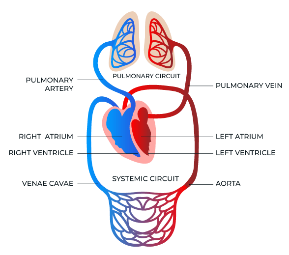
The pulmonary circuit (heart → lungs → heart)
This shorter circuit involves the right side of the heart. The right ventricle pumps deoxygenated blood to the lungs through the pulmonary artery. In the lungs, blood picks up oxygen and releases carbon dioxide. The now oxygenated blood returns to the left atrium through pulmonary veins. This circuit has relatively thin vessel walls because the distance is short and less pressure is needed.
The systemic circuit (heart → body → heart)
This longer circuit involves the left side of the heart. The left ventricle pumps oxygenated blood to all parts of the body through the aorta and its many branches. As blood travels through body tissues, it delivers oxygen and nutrients while collecting carbon dioxide and waste products. The deoxygenated blood then returns to the right atrium through the vena cava. This circuit requires higher pressure and thicker vessel walls because blood must reach every part of the body.
Benefits of the two-circuit system:
- Keeps oxygenated and deoxygenated blood completely separate
- Maintains high blood pressure throughout the body for efficient delivery
- Allows for more efficient gas exchange in the lungs
Cardiac blood supply
Even though the heart is constantly filled with blood, the heart muscle itself needs its own dedicated blood supply to function properly. This is because the blood inside the heart chambers cannot directly nourish the thick muscular walls.
Coronary arteries
The heart muscle receives oxygen and nutrients through a network of coronary arteries that branch from the aorta just as it leaves the heart. These arteries spread across the surface of the heart like a crown (which is what "coronary" means). The coronary arteries subdivide into numerous smaller vessels called cardiac capillaries that penetrate deep into the heart muscle.
Coronary veins
After delivering oxygen and nutrients, blood is collected by coronary veins that return the deoxygenated blood directly back to the right atrium through an opening called the coronary sinus.
Critical Information for Exams: Blockage of coronary arteries is a common cause of heart attacks. When coronary arteries become blocked, the heart muscle doesn't receive enough oxygen and begins to die. This is why maintaining healthy coronary arteries through good diet, exercise, and avoiding smoking is essential for heart health.
The pacemaker and heartbeat control
Natural pacemaker
The heart has its own internal timing system called the pacemaker or sinoatrial (SA) node. This small bundle of specialised tissue is located at the top of the right atrium and acts like the heart's natural battery. Even if the heart were removed from the body and kept in nutrient-rich fluid, it would continue beating because of this pacemaker.
How the pacemaker works
Step-by-Step: How Your Heartbeat is Controlled
Step 1: The SA node sends out an electrical signal automatically
Step 2: This signal causes both atria to contract simultaneously, pushing blood into the ventricles
Step 3: The signal reaches the atrioventricular (AV) node located between the atria and ventricles
Step 4: The AV node delays the signal briefly (allowing atria to empty completely)
Step 5: The signal travels down through special fibres in the septum
Step 6: Both ventricles contract together, but after the atria have finished contracting
This coordination ensures maximum pumping efficiency!
External control of heart rate
While the pacemaker sets the basic rhythm, the brain can influence heart rate through the nervous system and hormones. Factors that increase heart rate include exercise, excitement, stress, and temperature increases. Factors that decrease heart rate include relaxation, sleep, and certain chemicals like alcohol.
Heart sounds
When you listen to a heartbeat, you hear the characteristic "lub-dub" sound. These sounds are caused by the heart valves closing, not by the heart muscle contracting as many people think.
What causes heart sounds:
- The "lub" sound (first heart sound) occurs when the tricuspid and bicuspid valves slam shut as the ventricles begin to contract
- The "dub" sound (second heart sound) occurs when the semilunar valves close as the ventricles relax
Any unusual heart sounds, called heart murmurs, may indicate problems with one or more of the heart valves.
Key Points to Remember:
- The heart is a four-chambered muscular pump that beats about 100,000 times per day
- The septum divides the heart into left and right sides, keeping oxygenated and deoxygenated blood separate
- Valves ensure blood flows in only one direction: tricuspid and bicuspid valves between chambers, semilunar valves at exits
- The double circulation system has two circuits: pulmonary (to lungs) and systemic (to body)
- The heart's own blood supply comes from coronary arteries that branch from the aorta
- The pacemaker (SA node) controls heart rhythm automatically, but can be influenced by the nervous system
- Heart sounds ("lub-dub") are caused by valve closure, not muscle contraction