Human Reproduction (Grade 12 NSC Matric Life Sciences): Revision Notes
Implantation of the Blastocyst and Gestation
Understanding implantation
After fertilisation and early development, the blastocyst (a hollow ball of cells formed about 4 days after fertilisation) travels down the fallopian tube and enters the uterus. This is where the crucial process of implantation begins.
The implantation process happens in several important steps that allow the developing embryo to establish a connection with the mother's body. First, the blastocyst must find a suitable place to attach and begin growing.
The implantation process
When the blastocyst reaches the uterus, it doesn't simply stick to the wall - it actively creates its own space. The outer cells of the blastocyst release special enzymes that break down a small section of the thickened uterine wall (endometrium). This makes the uterine tissue softer and easier for the blastocyst to embed into.
The implantation process is active, not passive - the blastocyst actually secretes enzymes to prepare the endometrium for embedding. This shows how sophisticated even early development is!
Once the blastocyst sinks into this prepared area, something remarkable happens. The outer layers of cells begin developing into two essential membranes that will support the growing baby throughout pregnancy:
- Amnion - the inner membrane
- Chorion - the outer membrane
The chorion develops finger-like projections called chorionic villi that extend into the endometrium. These villi become part of the placenta and play a vital role in producing progesterone, a hormone essential for maintaining pregnancy.
At this point, we stop calling it a blastocyst and start using the term embryo.
The gestation period
Gestation is simply the scientific term for pregnancy - the time during which the embryo develops inside the mother's uterus. This period lasts approximately from the last menstrual period.
Key Timeline to Remember:
- 0-12 weeks: The developing baby is called an embryo
- 12+ weeks: The developing baby is called a foetus
This name change at 12 weeks reflects that all major organs and body systems have formed, and the focus shifts to growth and maturation.
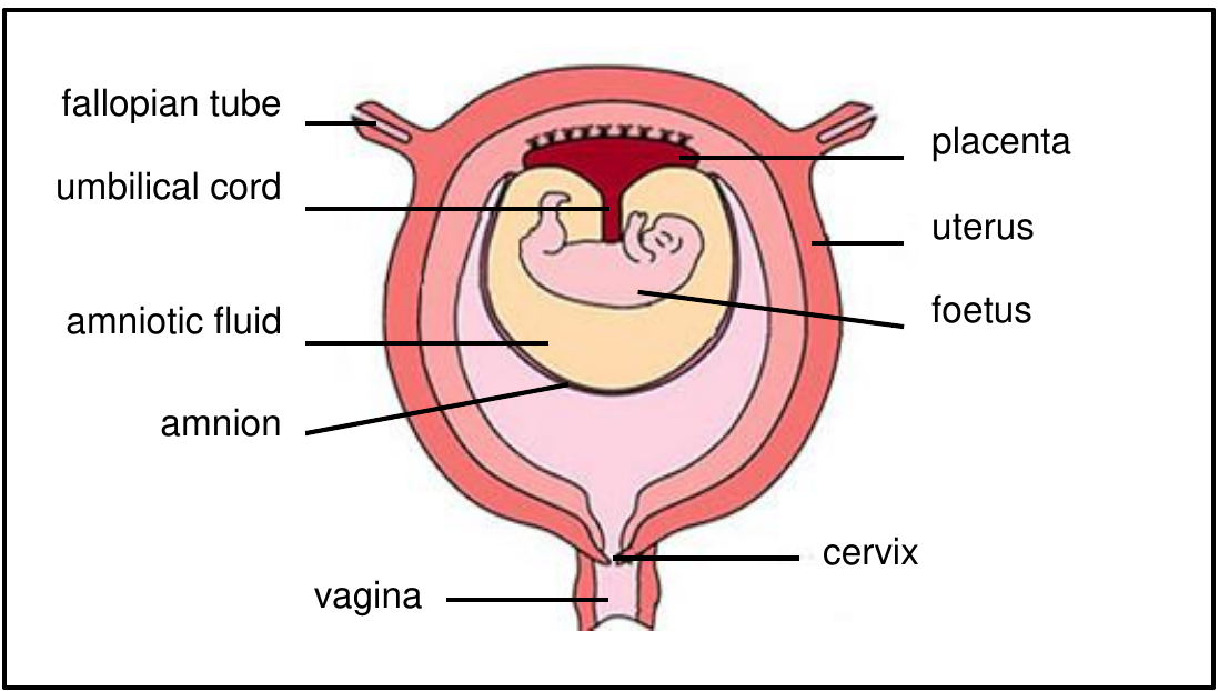
Extra-embryonic structures and their functions
The developing baby needs special support systems to survive and grow inside the mother's body. These structures form from the original blastocyst but exist outside the actual embryo/foetus itself.
The amnion and amniotic fluid
The amnion is the inner membrane that surrounds the developing baby. It produces amniotic fluid, which creates a protective environment with several crucial functions:
Functions of Amniotic Fluid:
Protection: Acts like a shock absorber, protecting the foetus from bumps and pressure changes
Temperature regulation: Keeps the foetus at a constant, ideal temperature
Movement: Allows the foetus to move freely, which is essential for proper muscle and bone development
Hydration: Prevents the foetus from becoming dehydrated
Think of amniotic fluid as a perfect cushioned environment - like being surrounded by warm, protective water.
The chorion and placenta formation
The chorion forms the outer membrane and plays a crucial role in creating the placenta. The chorionic villi that extend from the chorion help form the connection between mother and baby, allowing for the exchange of nutrients, oxygen, and waste products.
The umbilical cord
The umbilical cord is the vital lifeline connecting the foetus to the placenta. This cord contains three important blood vessels:
- Two umbilical arteries: These carry deoxygenated blood and waste products from the foetus to the placenta
- One umbilical vein: This carries oxygenated blood, nutrients, water, and other essential substances from the placenta to the foetus
In foetal circulation, the roles are opposite to normal circulation - arteries carry deoxygenated blood away from the foetus, while the vein brings oxygenated blood to the foetus. Remember: 2 arteries OUT, 1 vein IN.
The placenta - a remarkable temporary organ
The placenta is a temporary organ that forms where the blastocyst originally implanted. It serves as the interface between mother and baby, but here's something important to understand: the mother's blood and foetus's blood never actually mix.
Instead, substances pass between mother and foetus through a process called diffusion across the placental barrier.
Key functions of the placenta
The placenta serves several vital functions throughout pregnancy:
- Attachment point: Physically anchors the foetus to the mother's uterus
- Nutrient transfer: Allows nutrients from the mother's diet to reach the developing baby
- Gas exchange: Enables oxygen to pass from mother to foetus and carbon dioxide to pass from foetus to mother
- Waste removal: Allows waste products from foetal metabolism to be transferred to the mother for elimination
- Hormone production: After 12 weeks, the placenta takes over progesterone production to maintain the pregnancy
The placenta essentially acts as the foetus's lungs, kidneys, liver, and digestive system all rolled into one!
Common misconceptions and exam tips
Students often have difficulty with some key concepts in this topic, so it's worth clarifying these important points.
Common misconception: Many students think the mother's blood flows directly to the baby. This is incorrect - substances transfer by diffusion across the placental barrier, but the blood supplies remain separate.
When preparing for exams, focus on the key timeline - remember that gestation lasts 40 weeks, the embryo becomes a foetus at 12 weeks, and the placenta takes over hormone production at 12 weeks. When describing implantation, always mention the enzyme secretion that softens the endometrium - this shows understanding of the active process involved.
Key Points to Remember:
- Implantation is the process where the blastocyst embeds into the softened endometrium and develops into an embryo with supporting membranes
- Gestation lasts , with the embryo becoming a foetus after 12 weeks when all major organs have formed
- Amniotic fluid protects, regulates temperature, enables movement, and prevents dehydration of the developing foetus
- The umbilical cord contains 2 arteries (carrying waste away) and 1 vein (bringing nutrients and oxygen in)
- The placenta allows substance exchange without mixing maternal and foetal blood, serving as the foetus's life support system