Pathogens and Infectious Diseases: Further Exploration (HSC SSCE Biology): Revision Notes
Pathogens and Infectious Diseases: Further Exploration
Introduction to non-cellular pathogens
Non-cellular pathogens are infectious agents that lack cellular structure. Unlike bacteria, fungi, and protozoa, these pathogens cannot carry out metabolic processes independently. The two main types of non-cellular pathogens are viruses and prions. Understanding these unique disease-causing agents is crucial because they require different control strategies compared to cellular pathogens.
Non-cellular pathogens represent a unique category of infectious agents that challenge our traditional understanding of life. Their dependence on host cells makes them particularly difficult to treat without harming the host organism.
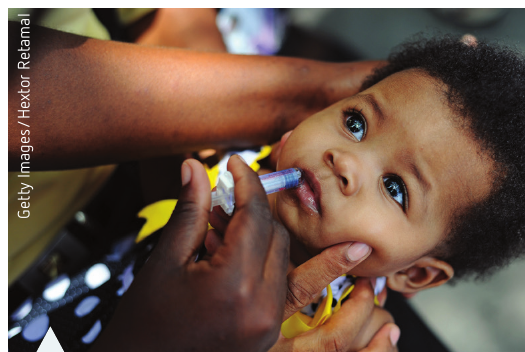
In Sydney during winter 2017, hospitals experienced a surge in patients with severe vomiting and diarrhoea. The outbreak particularly affected aged care facilities and childcare centres. The culprits were Norovirus and Rotavirus, both viral pathogens spread through direct contact with infected individuals. This real-world example demonstrates how viral infections can rapidly spread through vulnerable populations.
Viruses: characteristics and structure
What are viruses?
The word 'virus' comes from a Latin term meaning 'slimy liquid or poison'. Scientists first recognised viruses as pathogens in the 1890s when studying tobacco mosaic virus. Viruses occupy a unique position between living and non-living things, possessing characteristics of both.
Living characteristics:
- Contain genetic material (nucleic acids)
- Pass on hereditary information to offspring
- Can evolve and adapt
Non-living characteristics:
- Not composed of cells
- Cannot carry out metabolism independently
- Can be crystallised like chemicals
Viruses are obligate parasites, meaning they can only reproduce and metabolise inside a host cell. They are completely dependent on living cells for survival. A single viral particle is called a virion.
Virus structure
Viruses are extremely small, measuring only nm in diameter. They can only be viewed using an electron microscope. All viruses share a basic structural plan consisting of:
Core components:
- Genetic material: Either DNA or RNA (never both)
- Capsid: A protective protein coat surrounding the genetic material
- Envelope (in some viruses): A lipid membrane derived from the host cell that surrounds the capsid
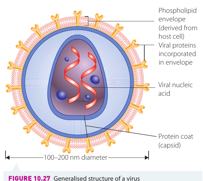
Viruses containing RNA as their genetic material are called retroviruses. The genetic material is the infectious part of the virus - this is what takes over the host cell's machinery.
Types of virus structures
Different viruses have distinct structural forms. The main types include:
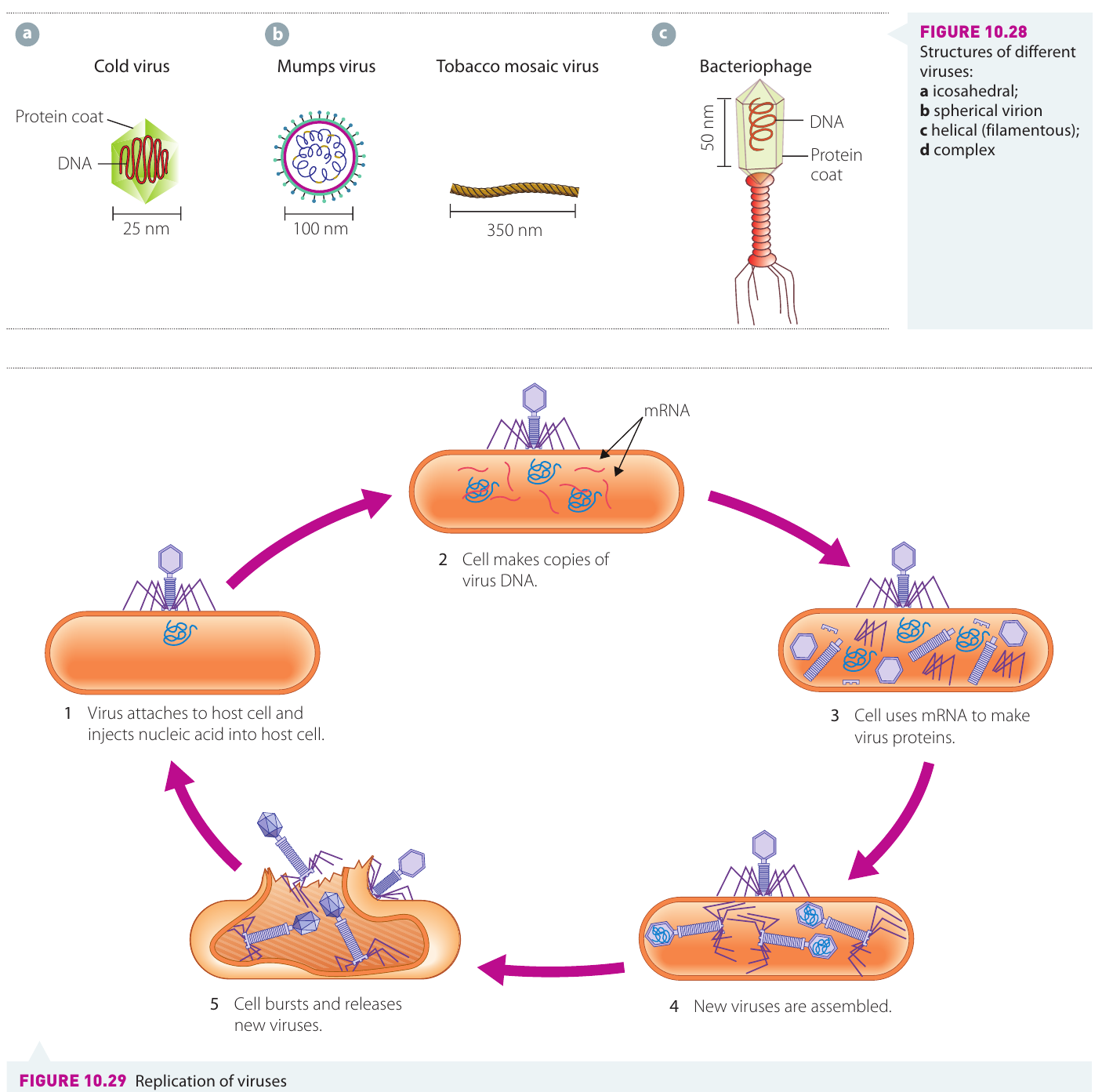
Icosahedral viruses (e.g., cold virus, nm): Twenty-sided symmetrical structure with a geometric appearance.
Spherical viruses (e.g., mumps virus, nm): Round shape with envelope surrounding the capsid.
Helical viruses (e.g., tobacco mosaic virus, nm): Rod-shaped or filamentous structure with genetic material arranged in a helix.
Complex viruses (e.g., bacteriophages, nm): Intricate structures with specialised parts like tail fibres and base plates.
How viruses replicate
Viruses cannot reproduce on their own. They hijack the host cell's reproductive mechanisms through a process called the lytic cycle:
The Viral Lytic Cycle: A Step-by-Step Process
Step 1 - Attachment and injection: The viral protein coat contains chemicals that allow the virus to attach to specific receptors on the host cell surface. Once attached, the virus either enters the cell whole or injects its genetic material inside.
Step 2 - DNA replication: The viral genetic material takes control of the host cell's machinery, forcing it to make copies of viral DNA or RNA.
Step 3 - Protein synthesis: The cell uses messenger RNA (mRNA) to produce viral proteins using the host's ribosomes.
Step 4 - Assembly: New virus particles are assembled inside the host cell from the newly synthesised viral components.
Step 5 - Release: The host cell becomes so full of new viruses that it dies and bursts (lysis), releasing hundreds or thousands of new virions that can infect other cells.
Bacteriophages (viruses that invade bacterial cells) use a slightly different approach. Instead of entering the host cell completely, they simply inject their genetic material through the cell wall while remaining attached to the outside.
Diseases caused by viruses
Viruses cause numerous diseases in humans, animals, and plants:
- Influenza (flu)
- Measles
- AIDS (caused by human immunodeficiency virus, HIV)
- Herpes
- Glandular fever
- SARS (severe acute respiratory syndrome)
- Tobacco mosaic virus disease in plants
Treatment challenges
Treating viral diseases is extremely difficult. Any substance that kills viruses will likely also damage or kill the host cells, because viruses rely entirely on host cell machinery. This is why antibiotics (which work against bacteria) are ineffective against viral infections. Treatment often focuses on managing symptoms while the immune system fights the infection, or using vaccines to prevent infection in the first place.
Prions: a unique pathogen
The discovery of kuru
Case Study: The Discovery of Kuru
In the highlands of Papua New Guinea during the 1950s, researchers discovered an unusual disease among the Fore people. The disease, called kuru, caused shivering, trembling, and often uncontrolled laughter, progressing rapidly to death. At least 200 people per year died from this mysterious condition.
Researchers eventually linked the disease to mortuary practices. When someone died, their brain was consumed by relatives to spare them from being eaten by worms. This practice transmitted the infectious agent. The scientists who identified this completely new type of pathogen - the prion - won a Nobel Prize in Physiology or Medicine for their groundbreaking work.
What are prions?
A pathogenic prion is an abnormal protein capable of causing degenerative diseases of the nervous system. Prions are fundamentally different from all other pathogens:
- No genetic material: Unlike viruses, bacteria, fungi, and protozoa, prions contain no DNA or RNA
- Smallest pathogens: Even smaller than viruses
- Pure protein: Consist only of misfolded protein molecules
Normal prion proteins are found naturally in the brain and spinal cord of humans and mammals, where they perform helpful functions. However, pathogenic prions have a different three-dimensional shape.
Prion structure and mechanism
The key difference between normal and pathogenic prions lies in their protein folding:
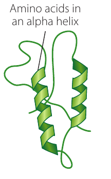
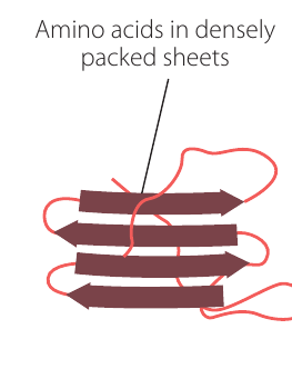
Normal prion protein: Contains amino acids arranged in alpha helix structures - spiral, coiled shapes that are loosely packed.
Infectious prion protein: Contains amino acids arranged in beta-pleated sheets - flat, densely packed layers stacked together.
How prions cause disease
Pathogenic prions cause disease through a domino-like effect. When an infectious prion comes into contact with a normal prion protein, it induces the normal protein to change its shape and become infectious too. This creates a chain reaction:
- Infectious prion encounters normal prion protein
- Normal protein refolds into the pathogenic shape
- Both proteins are now infectious
- Each infectious prion converts more normal proteins
- Abnormal proteins accumulate in nervous tissue
The accumulating abnormal proteins are deposited within the central nervous system and other organs, causing progressive damage.
Effects on brain tissue
Prion diseases are called transmissible spongiform encephalopathies (TSEs) because the brain tissue of infected individuals becomes full of holes, resembling a sponge:
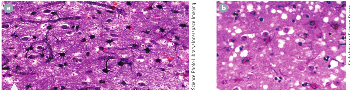
The images show normal brain tissue compared to tissue affected by prion disease. The white spaces represent holes where brain tissue has been destroyed by accumulating abnormal prions.
Characteristics of TSE diseases
All prion diseases share certain features:
- Very long incubation period: Typically years between infection and first symptoms
- Rapid progression: Once clinical signs appear, the disease advances quickly
- Always fatal: No treatments can cure or stop prion diseases
- Degenerative: Progressive deterioration of nervous system function
How prions are transmitted
Prion diseases can be contracted through several routes:
Dietary exposure:
- Ingesting tissue containing infectious prions, particularly nervous tissue and brain
- Eating contaminated meat products
Medical procedures:
- Surgery using contaminated equipment that was not properly sterilised
- Receiving contaminated growth hormone injections from infected animals
- Receiving corneal transplants from previously infected organ donors
Genetic transmission:
- Inheriting the mutated gene that codes for the infectious prion form
Spontaneous formation:
- Random conversion of normal prion proteins to infectious forms (very rare)
Diseases caused by prions
Important prion diseases include:
In humans:
- Creutzfeldt-Jakob disease (CJD): Rapidly progressive and fatal brain disease
- Variant CJD: Linked to consuming beef from cattle with BSE ('mad cow disease')
- Kuru: Transmitted through mortuary cannibalism in Papua New Guinea
- Fatal familial insomnia: Spontaneous transformation of protein in the brain
In animals:
- Bovine spongiform encephalopathy (BSE): 'Mad cow disease' in cattle
- Chronic wasting disease: Affects deer, elk, and moose in North America
- Scrapie: Affects sheep and goats
Modes of transmission of infectious disease
Understanding how diseases spread is critical for controlling outbreaks. Transmission involves carrying or transferring a pathogen from an infected host to a non-infected organism.
Pathogen reservoirs and carriers
Pathogens often exist in reservoirs - environments or living hosts where they can survive outside an active infection. The ability of a pathogen to survive outside a host relates directly to its transmission mode.
Environmental reservoirs:
- Spore-forming bacteria like anthrax can exist for years in soil where infected animal fluids have contaminated the ground
- Water sources can harbour various pathogens
- Contaminated surfaces and objects
Living reservoirs:
- The human gut can harbour disease-causing organisms
- Animals can carry diseases that affect humans
- Intermediate hosts in parasite life cycles
Human carriers:
Active carriers harbour the disease in their own body. They may or may not show symptoms. A famous example is Mary Mallon ('typhoid Mary'), an asymptomatic carrier of typhoid fever (Salmonella typhi). Working as a cook in New York City between 1900 and 1915, she infected around 122 people, five of whom died.
Passive carriers transmit pathogens from person to person without being infected themselves. This commonly occurs in healthcare settings when staff fail to wash hands between patients.
Complex life cycles in parasites
Macroparasites often have complex life cycles involving multiple hosts and transmission stages. The parasite must successfully complete each stage and be transmitted between hosts to reach sexual maturity.
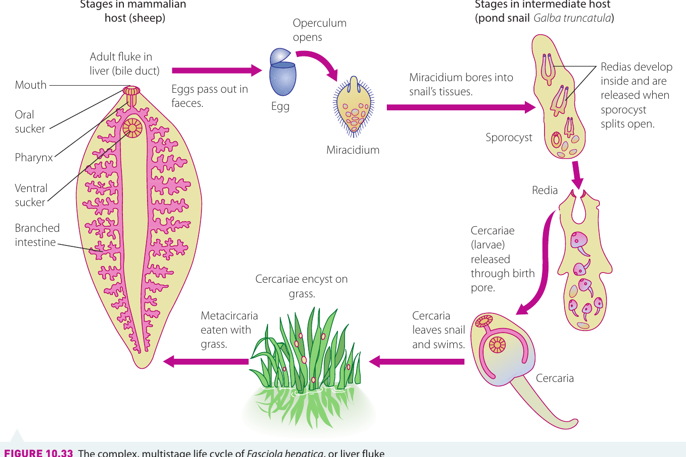
The Liver Fluke Life Cycle: A Complex Journey
The liver fluke (Fasciola hepatica) demonstrates this complexity. Its life cycle involves:
Primary host (sheep/cattle):
- Adult flukes live in the liver bile duct
- Eggs pass out in faeces
Free-living stage: 3. Eggs develop in water and release miracidia (larvae)
Intermediate host (pond snail): 4. Miracidium penetrates snail tissue 5. Develops into sporocyst 6. Produces rediae 7. Rediae produce cercariae (swimming larvae) 8. Cercariae leave the snail
Environmental stage: 9. Cercariae encyst on grass as metacercariae
Back to primary host: 10. Grazing animals eat grass with metacercariae 11. Larvae develop into adult flukes in the liver
This complex cycle requires multiple successful transmissions for the parasite to complete its life cycle.
The chain of infection
For a disease to spread between organisms, a 'chain of infection' must exist. This chain has three essential elements:
- A susceptible host: An organism that can be infected by the disease
- A pathogen: An organism capable of causing the disease
- A mode of transmission: A way for the pathogen to travel from host to host
Breaking any link in this chain prevents disease transmission. This principle underlies many disease control strategies.
Three modes of transmission
All disease transmission falls into three main categories:
Direct contact - Transfer through physical contact with infected skin or body secretions
Indirect contact - Transfer through non-living objects or environmental sources
Vector transmission - Transfer through another organism, typically an arthropod
Direct contact transmission
Direct contact transmission occurs when there is physical contact between an infected host and a non-infected organism. This transmission can be classified as:
Horizontal transmission: Contact between organisms of the same generation, or between organisms that are not parent and child.
Vertical transmission: Contact between offspring and parent, such as from mother to baby during childbirth.
Types of direct contact
Physical contact that can transmit disease includes:
- Touching infected skin
- Sexual contact
- Kissing
- Contact with nasal or oral secretions
- Biting
- Direct contact with blood or other body fluids
- Direct contact with open wounds
- Prenatal transmission: Before birth or during pregnancy
- Perinatal transmission: Around the time of birth, including during delivery
Diseases spread by direct contact
Common examples include:
- Skin infections: Ringworm and impetigo spread through touching infected skin
- Cytomegalovirus (CMV): Transmitted through saliva and other body fluids
- Glandular fever: Spread through saliva (the 'kissing disease')
- HIV/AIDS: Transmitted through blood and sexual contact
- Herpes simplex virus (HSV): Spread through direct contact with infected areas
Indirect contact transmission
Indirect contact transmission occurs when the host and another organism have no direct physical contact. Instead, infection occurs from a reservoir created by the host outside itself.
Types of indirect transmission
Airborne transmission:
- Coughing or sneezing releases droplets containing pathogens
- These droplets can travel up to metres through the air
- Other people inhale the contaminated droplets
Surface contamination:
- Touching infected surfaces or objects
- A fomite is any object or substance that carries infection
- Examples include door handles, utensils, towels, and medical equipment
Contaminated food or water:
- Pathogens survive in food or water sources
- Consumption leads to infection
Medical equipment:
- Infected surgical instruments not properly sterilised
- Surgical instruments are generally sterilised using saturated steam under pressure in an autoclave
Vector transmission:
- Discussed in detail in the next section
Diseases spread by indirect contact
Important examples include:
- Measles virus: Spreads through infected droplets in the air
- Gastroenteritis: E. coli bacteria transmitted through contaminated food and water
- Toxoplasmosis: Protozoan Toxoplasma gondii spread through infected cat droppings
- Legionnaires' disease: Over 50 species of Legionella bacterium transmitted through contaminated water in cooling towers
- Influenza: Exposure to droplets and biological matter containing virus particles
Airborne diseases pose particular challenges because they are often the most difficult to control once an outbreak occurs. People can be infected without any direct contact with sick individuals.
Vector transmission
Vector transmission is a specialised form of indirect transmission involving living organisms that carry pathogens from one host to another.
Common vectors
Arthropod vectors:
- Certain species of mosquitoes
- Sandflies
- Ticks
- Fleas
- Flies
Other vectors:
- Infected aquatic snails
- Mammals such as fruit bats (Hendra virus) and pigs (Menangle virus)
- Sometimes infected plants and fungi
How vector transmission works
Most vector transmission involves arthropods that feed on blood. The typical process is:
- Vector bites or feeds on infected host
- Pathogen enters vector's body
- Pathogen may or may not replicate in the vector
- Vector bites new host
- Pathogen is transmitted during feeding
In some cases, animals swallow the arthropod vector during grooming (for example, bot flies in horses, or flea tapeworms in dogs).
Environmental and social factors
Vector-borne diseases represent around of all infectious diseases in humans. Every year, millions of people die from infections transmitted by vectors.
Several factors influence the transmission of these diseases:
Environmental factors:
- Vector diseases are most common in warm, humid climates
- These conditions favour insect survival and reproduction
- Female Anopheles mosquitoes (malaria vectors) require water to lay eggs
- Climate change may expand the range of disease vectors
Social factors:
- Housing quality affects exposure to vectors
- Access to screens, insect repellents, and bed nets
- Public health infrastructure and disease surveillance
- Education about disease prevention
Diseases spread by vectors
Important vector-borne diseases include:
- Chagas disease: Transmitted by triatomine bugs
- Malaria: Spread by female Anopheles mosquitoes
- Dengue fever: Transmitted by Aedes mosquitoes
- Leishmaniasis: Spread by sandflies (now found in Australian macropods)
- Schistosomiasis: Transmitted by aquatic snails
- Onchocerchiasis: Spread by blackflies
- Canine and feline heartworm: Dirofilaria immitis transmitted by mosquitoes
- Hendra and Nipah viruses: Carried by fruit bats
Case study: equine influenza virus outbreak
This case study demonstrates how understanding transmission is critical for disease control.
Background
Equine influenza (horse flu) is an exotic disease in Australia - it is not normally found here. If it became enzootic (endemic in an animal population), it would devastate the Australian horse industry. The 2007 outbreak demonstrates the importance of rapid response and biosecurity measures.
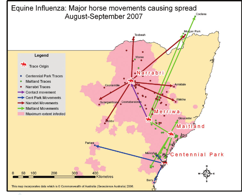
The 2007 Australian Equine Influenza Outbreak
In August 2007, an equine veterinarian reported several sick horses in Centennial Park, Sydney. Simultaneously, breeding stallions from Japan at the Eastern Creek Quarantine Station showed signs of equine influenza virus (EIV) infection after importation. Laboratory tests confirmed an outbreak in both groups. Protocols established in the Australian Veterinary Emergency Plan (AUSTVETPLAN) were immediately activated.
Cause and symptoms
Pathogen: Equine influenza virus is an orthomyxovirus affecting horses and donkeys. It does not infect humans.
Virus strains:
- Equine-1 (H7N7)
- Equine-2 (H3N8)
Clinical signs:
- Fever ( or higher)
- Watery nasal discharge
- Hacking cough
- Loss of appetite
- Muscle pain
- Depression
- Laboured breathing
Transmission pathways
Equine influenza is highly contagious and spreads through two main routes:
Direct transmission:
- Between infected horses through nasal secretions
- Contact with other body fluids from infected animals
Indirect transmission:
- Through humans carrying the virus on contaminated clothing, shoes, or equipment
- Via contaminated grooming tools
- Through shared food and water buckets
- On contaminated vehicles and transport equipment
Management of the outbreak
Once diagnosis was confirmed, rapid action prevented catastrophic spread:
Immediate response:
Movement restrictions: The NSW Chief Veterinary Officer imposed a state-wide lockdown on all horse movements, which eventually became nation-wide. This prevented further spread of the virus to unaffected areas.
Coordination centre: A management centre coordinated activities at disease control headquarters in Orange and Menangle, NSW.
Quarantine measures: Horse properties throughout NSW were quarantined, preventing any animals from leaving or entering.
Disease mapping: The spread was carefully tracked. By late August, the virus had reached the NSW Central Coast and Hunter Valley. Areas with high horse populations (such as Dubbo) experienced the fastest disease spread.
Outbreak trajectory
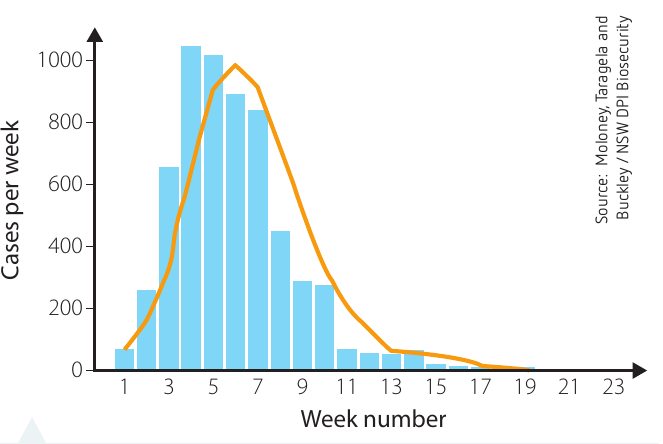
The epidemic curve shows the pattern of new cases over time:
- Cases increased rapidly in the first few weeks
- Peak incidence occurred around weeks with approximately 1000 cases per week
- Numbers declined steadily after the peak
- By week 15, very few new cases were reported
With the lockdown of horse movements and strict quarantine procedures, the outbreak was controlled by December 2007. Australia was declared free of the virus in early 2008. This successful eradication prevented the virus from becoming established in the Australian horse population.
Control of future outbreaks
The Australian Veterinary Association concluded that mass vaccination is not a justifiable option for several reasons:
Why vaccination is not recommended:
- The equine influenza virus mutates similarly to human influenza viruses
- Vaccination would not provide immunity against new strains
- Even vaccinated horses can spread the virus indirectly
- Vaccination might delay disease detection by masking symptoms
Recommended control measures:
Import restrictions:
- Limit importation of live horses to those from approved countries only
- Assess disease status in exporting countries regularly
Strict biosecurity measures:
- Quarantine imported horses in their country of origin for 14 days before export
- Quarantine all arrivals in post-entry facilities in Victoria for minimum 14 days
- Monitor horses closely for any signs of illness during quarantine
Public education:
- Particularly target those working in the horse industry
- Early detection depends on recognising symptoms quickly
- Educate about biosecurity practices
Biosecurity training:
- Train all personnel involved in horse importation
- Include grooms, truck drivers, cleaners, and airline staff
- Emphasise hygiene and disease prevention protocols
This case study illustrates how understanding transmission pathways allows for effective disease control through breaking the chain of infection.
Identifying microbes in food and water
Microbes are present everywhere in our environment - on our bodies, in the air, on surfaces, in water, and in food. While most microbes are harmless or even beneficial, some can contaminate food and water, causing illness.
Detection challenges and solutions
Individual microbes are too small to see with the naked eye. However, when provided with suitable conditions for reproduction and growth, many microbes cluster together, forming visible colonies.
Conditions needed for colony growth:
- Moisture
- Nutrients
- Warmth (typically around )
Laboratory methods:
Scientists use nutrient agar plates to grow microbial colonies. Agar is a jelly-like substance derived from seaweed. It is dissolved in water, mixed with nutrients suitable for microbial growth, and allowed to set in shallow dishes called Petri dishes.
Different pathogens require different nutrients. For example, streptococci bacteria often need nutrient medium enriched with sheep blood to grow well. Samples from food or water are spread onto the agar surface, then the plates are incubated at approximately to encourage growth.
Colony characteristics
After incubation, visible colonies appear on the agar surface. These colonies can be identified based on distinctive features:
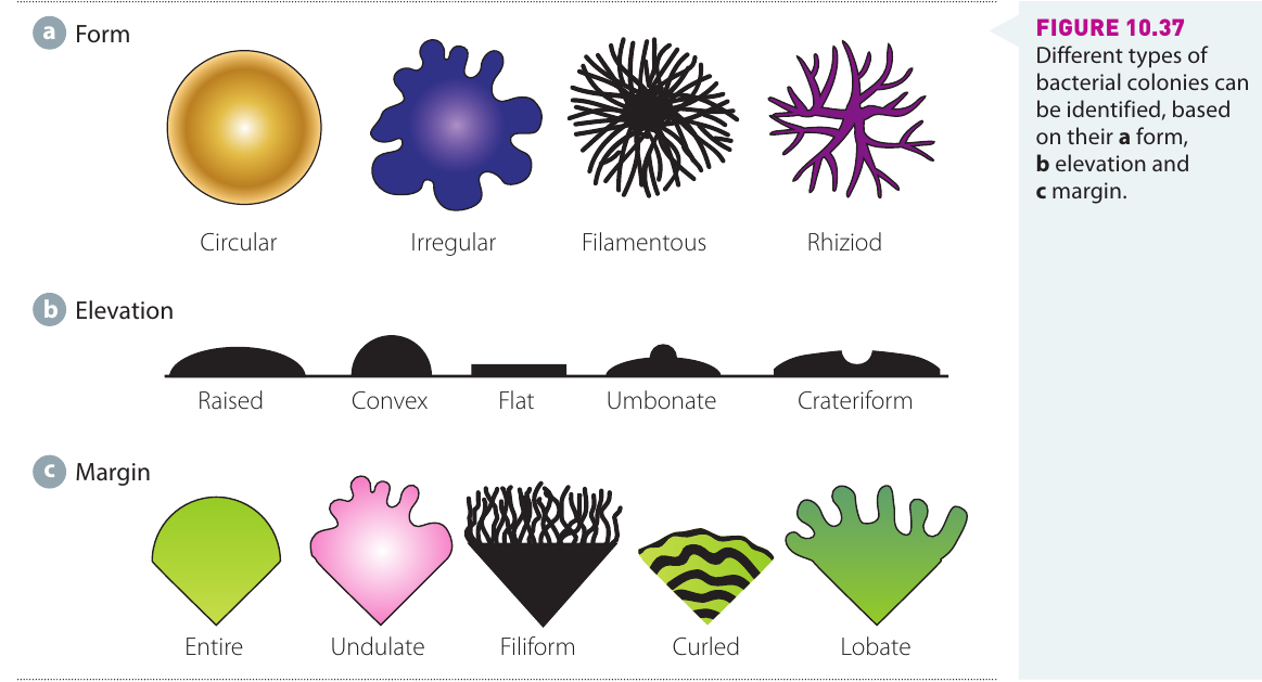
Form (basic shape):
- Circular: Round, even edge
- Irregular: Uneven, asymmetric shape
- Filamentous: Thread-like projections extending outward
- Rhizoid: Root-like, branching appearance
Elevation (cross-sectional shape):
- Raised: Slightly elevated above agar surface
- Convex: Dome-shaped, rounded top
- Flat: Level with agar surface
- Umbonate: Raised centre with flatter edges
- Crateriform: Depressed centre, crater-like
Margin (edge pattern):
- Entire: Smooth, even edge
- Undulate: Wavy edge
- Filiform: Thread-like projections at edge
- Curled: Curved, spiral edge pattern
- Lobate: Lobed, finger-like projections
Additional features:
- Colour: Varies widely - white, yellow, orange, pink, or other colours
- Surface texture: Smooth, dull, shiny, wrinkled, or rough
- Size: Diameter of the colony
Distinguishing bacterial and fungal colonies
Bacterial colonies typically appear:
- Smooth and glossy
- Coloured (various colours)
- Relatively small
- With defined edges
Fungal colonies typically appear:
- Furry or fuzzy
- Large
- Often white or grey
- With spreading, diffuse edges
By examining these characteristics, microbiologists can identify the types of microbes present in food or water samples, helping to ensure safety and detect contamination.
Remember!
Key Points to Remember:
-
Viruses are non-cellular pathogens containing DNA or RNA in a protein coat. They are obligate parasites that can only reproduce inside host cells by hijacking cellular machinery.
-
Prions are abnormal proteins without any genetic material. They cause disease by inducing normal prion proteins to misfold into the pathogenic form, creating a chain reaction. Prion diseases (TSEs) have long incubation periods but are always fatal.
-
The chain of infection requires three elements: a susceptible host, a pathogen capable of causing disease, and a mode of transmission. Breaking any link stops disease spread.
-
Three modes of transmission exist: direct contact (physical contact with infected individuals), indirect contact (through contaminated objects, surfaces, food, water, or air), and vector transmission (through organisms like mosquitoes or ticks).
-
Understanding transmission pathways is essential for disease control, as demonstrated by the successful management of the 2007 equine influenza outbreak in Australia through movement restrictions, quarantine, and biosecurity measures.