Human Immunodeficiency Virus (HIV) (AQA A-Level Biology): Revision Notes
Human Immunodeficiency Virus (HIV)
Introduction to HIV
Human Immunodeficiency Virus (HIV) is the causative agent of Acquired Immune Deficiency Syndrome (AIDS). HIV represents a relatively recent pathogen, first identified in 1981, and has since become a major global health concern.
HIV belongs to a group of viruses called retroviruses. These are characterised by their ability to synthesise DNA from RNA using the enzyme reverse transcriptase - the opposite process to normal transcription.
The ability of retroviruses to convert RNA to DNA is unique in biology and represents a reversal of the normal flow of genetic information (DNA → RNA → protein). This unusual characteristic is what makes HIV particularly challenging to treat and eradicate from infected individuals.
Structure of HIV
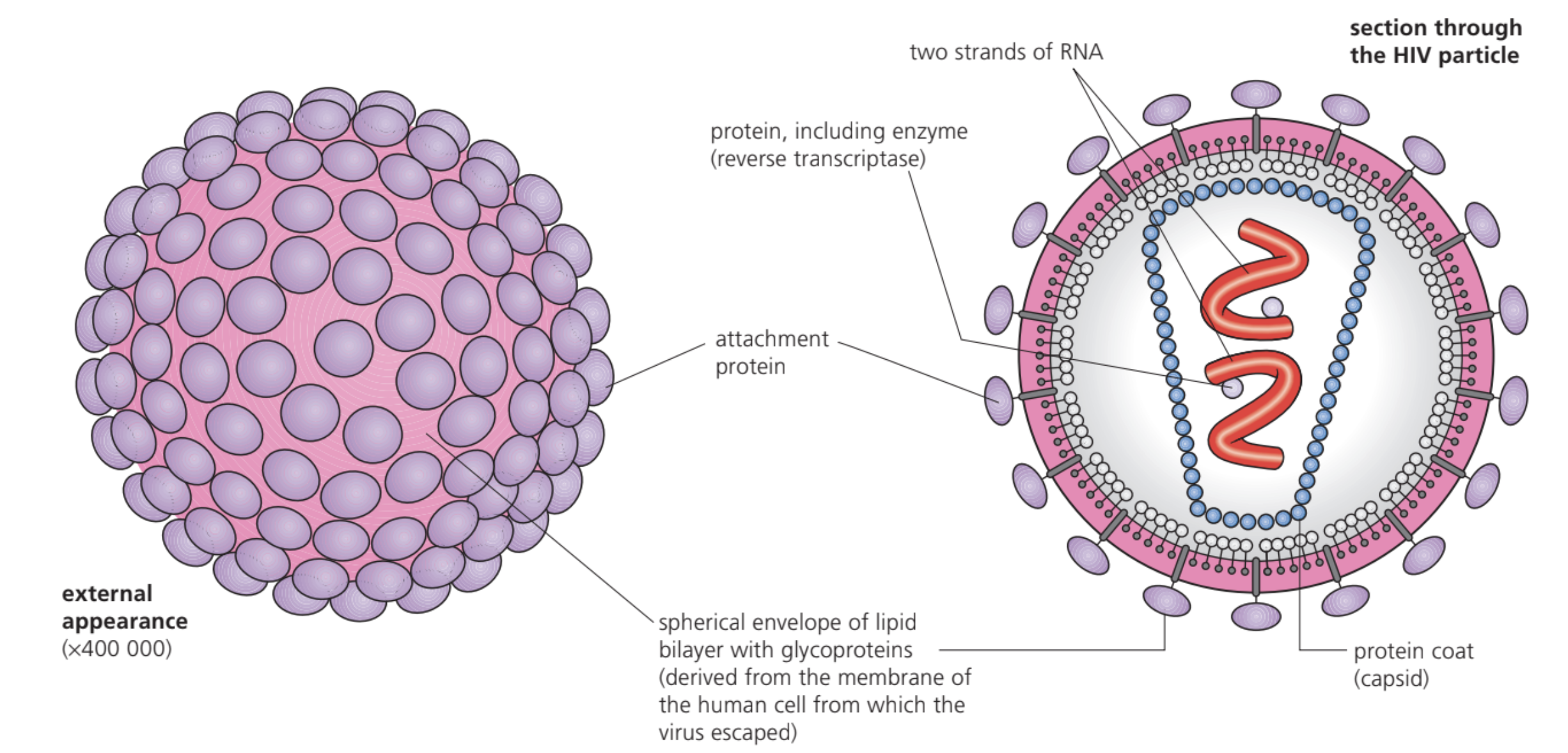
HIV exhibits a complex viral structure with several distinct components that work together to enable successful infection and replication:
- Lipid envelope: The outer layer consists of a lipid membrane derived from the host cell. Embedded within this envelope are attachment proteins that appear as spike-like projections on the viral surface.
- Capsid: Inside the lipid envelope lies a protein shell that protects the viral genetic material and enzymes.
- Genetic material: HIV contains two single strands of RNA rather than DNA, which is characteristic of retroviruses.
- Enzymes: The virus carries several enzymes within its structure, most notably reverse transcriptase. This enzyme enables HIV to convert its RNA genome into DNA, allowing integration into the host cell's genetic material.
The unique structure of HIV makes it well-adapted for infecting specific immune cells and establishing persistent infections.
The lipid envelope of HIV is actually derived from the membrane of the previous host cell it infected. This means that each new viral particle carries a piece of its former host, which helps it evade immune detection and facilitates infection of new cells.
Replication of HIV
As a virus, HIV cannot replicate independently and must hijack host cell machinery. Understanding this complex process is crucial to comprehending how HIV establishes infection and persists in the human body.
Step-by-Step HIV Replication Process
Step 1 - Initial infection: HIV enters the bloodstream and circulates throughout the body.
Step 2 - Cell binding: The virus specifically targets cells expressing the CD4 protein on their surface. While this protein appears on various cell types, HIV most commonly infects helper T cells.
Step 3 - Membrane fusion: The viral capsid fuses with the host cell membrane, allowing the viral RNA and enzymes to enter the helper T cell.
Step 4 - Reverse transcription: Once inside the cell, reverse transcriptase converts the viral RNA into DNA - a process unique to retroviruses.
Step 5 - Integration: The newly synthesised viral DNA moves into the cell's nucleus where it becomes incorporated into the host cell's chromosomal DNA.
Step 6 - Transcription and translation: The integrated viral DNA instructs the host cell to produce messenger RNA (mRNA) using the cell's own enzymes. This mRNA contains instructions for manufacturing new viral proteins and RNA.
Step 7 - Viral assembly: The mRNA moves out through nuclear pores and uses the host cell's protein synthesis machinery to produce HIV components.
Step 8 - Viral release: New HIV particles are assembled and break away from the helper T cell, acquiring a portion of the cell membrane which forms their lipid envelope.
Following infection, a person becomes HIV positive. However, the virus often enters a dormant phase and may not progress to AIDS until many years later.
Once HIV DNA integrates into the host cell's chromosomes, it becomes a permanent part of that cell's genetic material. This integration makes HIV extremely difficult to eliminate from the body, even with current treatments, as the viral DNA can remain dormant and reactivate later.
How HIV causes AIDS
HIV specifically targets and destroys helper T cells, leading to progressive immune system failure. This process represents one of the most devastating effects of viral infection on human health.
The destruction occurs through several interconnected mechanisms:
- Direct cell destruction: HIV kills helper T cells or interferes with their normal functioning.
- Immune system collapse: A healthy individual typically maintains between 800-1200 helper T cells per cubic millimetre of blood. In AIDS patients, this count can drop to as low as 200 per cubic millimetre.
- Loss of immune coordination: Helper T cells play essential roles in cell-mediated immunity. Without sufficient numbers, the immune system cannot effectively stimulate B cells to produce antibodies or activate cytotoxic T cells to eliminate infected cells.
Critical Immune System Failure
The helper T cell count is crucial for diagnosis and monitoring HIV progression. When counts fall below 200 cells per mm³, patients are considered to have progressed to AIDS, regardless of symptoms. At this stage, the immune system becomes critically compromised and unable to protect against even normally harmless organisms.
- Secondary infections: The compromised immune system becomes unable to mount adequate responses against other pathogens. AIDS patients frequently develop infections affecting the lungs, intestines, brain, and eyes, along with experiencing weight loss and diarrhoea.
- Opportunistic cancers: The weakened immune surveillance allows certain cancers to develop more readily.
HIV does not directly kill individuals. Instead, it prevents normal immune function, making patients vulnerable to secondary infections and diseases that ultimately prove fatal.
The ELISA test
ELISA (Enzyme Linked Immunosorbent Assay) provides a highly sensitive method for detecting and quantifying proteins, including HIV antigens. The test can identify extremely small amounts of target molecules, making it invaluable in medical diagnostics.
ELISA Testing Procedure
Step 1 - Sample application: The biological sample is applied to a surface (typically a slide or microwell) where target antigens will bind.
Step 2 - Initial washing: The surface undergoes thorough washing to remove unattached antigens.
Step 3 - Primary antibody addition: Specific antibodies that recognise the target antigen are added and allowed to bind.
Step 4 - Second washing: Excess unbound primary antibodies are removed through washing.
Step 5 - Secondary antibody addition: A second antibody that binds to the first antibody is introduced. This secondary antibody has an enzyme attached to it.
Step 6 - Substrate addition: A colourless substrate for the enzyme is added. The enzyme converts this substrate into a coloured product.
Step 7 - Quantification: The intensity of colour development correlates directly with the amount of target antigen present in the original sample.
ELISA proves particularly valuable for detecting HIV and other disease-causing organisms including tuberculosis and hepatitis. The technique's high sensitivity makes it excellent for measuring drug concentrations and conducting allergen testing.
The colour intensity in ELISA is directly proportional to the amount of target antigen present. This quantitative aspect makes ELISA not only useful for detecting the presence of HIV but also for monitoring viral load levels in patients undergoing treatment.
Why antibiotics are ineffective against viruses
Antibiotics work by targeting specific structures or processes in bacterial cells, making them completely ineffective against viral infections like AIDS. Understanding this difference is crucial for proper medical treatment approaches.
Bacterial cell wall disruption: Many antibiotics, such as penicillin, inhibit enzymes required for synthesising and assembling murein (peptidoglycan) cross-linkages in bacterial cell walls. This weakens the cell wall structure.
Osmotic pressure and cell death: Bacterial cells constantly face osmotic pressure as water enters through their permeable membranes. The strong murein cell wall normally prevents cell bursting. When antibiotics compromise this wall, water entry causes the bacterial cell to swell and eventually burst.
Key Structural Differences
Viruses possess a protein coat rather than a murein cell wall. They lack the cellular structures and metabolic pathways that antibiotics target. Additionally:
- Viruses depend entirely on host cells for their metabolic activities and replication
- They have no independent metabolic mechanisms that antibiotics can disrupt
- When viruses reside within host cells, antibiotics cannot reach them effectively
This fundamental difference in structure and biology explains why viral infections require different therapeutic approaches compared to bacterial diseases.
Intracellular location: When viruses reside within host cells, antibiotics cannot reach them effectively, further limiting treatment options.
This structural and biological distinction emphasises why antiviral medications rather than antibiotics are required for treating HIV and other viral infections.
Summary
Key Points to Remember:
-
HIV is a retrovirus that uses reverse transcriptase to convert RNA into DNA, allowing integration into host cell chromosomes
-
The virus specifically targets helper T cells by binding to CD4 proteins, leading to progressive immune system destruction
-
AIDS results from secondary infections and cancers that develop when helper T cell counts fall critically low (below 200 per mm³)
-
ELISA testing provides highly sensitive detection of HIV antigens using enzyme-linked antibodies and colourimetric reactions
-
Antibiotics cannot treat viral infections because viruses lack cell walls and independent metabolic pathways that these drugs target