Methods of Studying Cells (AQA A-Level Biology): Revision Notes
Microscope Measurements & Calculations
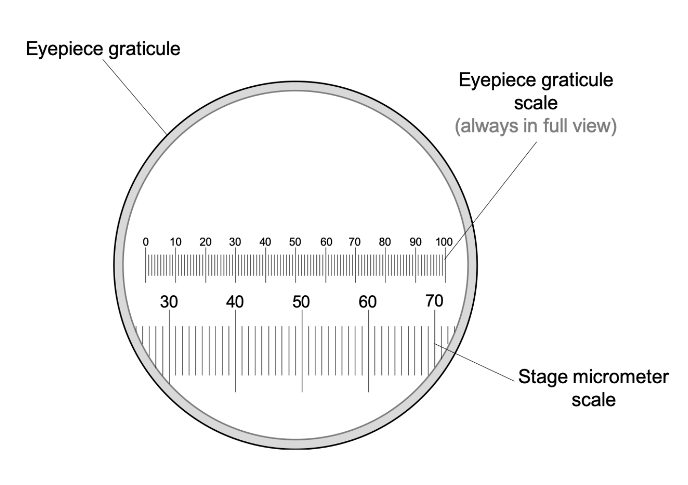
When studying cells under a light microscope, scientists need accurate methods to determine the size of cellular structures and organisms. The eyepiece graticule provides this measurement capability. This device consists of a glass disc positioned within the microscope's eyepiece, featuring an etched scale that appears as a ruler when viewing specimens.
A standard eyepiece graticule measures approximately 10mm in total length and contains 100 individual divisions marked along its scale. However, the graticule scale cannot directly measure specimen sizes because different objective lenses produce varying levels of magnification, making each division represent different actual distances depending on which lens is being used.
The eyepiece graticule appears as a fixed scale in your field of view, but its actual measurement value changes depending on the objective lens magnification. This is why calibration is essential for accurate measurements.
Calibrating the eyepiece graticule
Why calibration is essential
Since each objective lens magnifies specimens to different degrees, the eyepiece graticule must be calibrated for each specific objective lens before accurate measurements can be taken. This calibration process establishes the relationship between graticule divisions and real distances, allowing the same graticule to remain useful across different magnifications.
Critical Point: Each objective lens requires separate calibration. Never assume that a calibration value determined for one objective lens will work for another magnification level.
The calibration process
Calibration requires a specialised slide called a stage micrometre. This precision instrument features an etched scale typically measuring 2mm in total length, with the smallest divisions representing 0.01mm (equivalent to 10μm). The stage micrometre provides the known reference measurements needed for calibration.
Stage micrometres are precisely manufactured instruments with known, accurate measurements. They serve as the "ruler" that allows you to determine what each graticule division actually represents in real distance.
To calibrate an eyepiece graticule:
- Place the stage micrometre on the microscope stage
- Align the scales of both the eyepiece graticule and stage micrometre
- Count how many graticule divisions correspond to a known number of micrometre divisions
- Calculate the value of each graticule division for that specific objective lens
Calibration calculations
The calibration process involves establishing ratios between the two scales.
Worked Example: Basic Calibration
If 10 units on the stage micrometre scale align with 40 units on the graticule scale, then:
Step 1: Set up the ratio 10 micrometre units = 40 graticule units
Step 2: Find the relationship 1 micrometre unit = 4 graticule units
Step 3: Calculate graticule unit value Since each micrometre unit represents 10μm: Each graticule unit = 10μm ÷ 4 = 2.5μm
For different objective lenses, you can calculate new calibrations by considering magnification differences. If a ×40 objective gives a calibration of 2.5μm per graticule unit, then a ×400 objective (10 times greater magnification) would give 2.5μm ÷ 10 = 0.25μm per graticule unit.
Calculating linear magnifications from images
Understanding magnification calculations
When examining photographs or drawings of microscopic specimens, you can determine the linear magnification using the relationship between the image size and the actual specimen size. This calculation helps verify the scale of microscopic images and understand the degree of enlargement.
The magnification formula
Unit Consistency: Always ensure both measurements are in the same units before calculating. Convert millimetres to micrometres (1mm = 1,000μm) or vice versa as needed.
Working through magnification examples
Consider a photograph showing a cellular structure where a line marked X—Y represents a 5μm length in the actual cell. This type of measurement is common when analysing microscopic images.
Worked Example: Calculating Magnification
If the line X—Y measures 23mm on the photograph:
Step 1: Convert units to be consistent
- Actual size: 5μm
- Image size: 23mm = 23,000μm
Step 2: Apply the magnification formula
Result: The photograph shows the specimen enlarged 4,600 times compared to its real size.
Calculating actual sizes from images
Determining real specimen dimensions
When you know both the magnification and the measured size in an image, you can work backwards to find the actual size of cellular structures. This reverse calculation proves particularly useful when analysing electron micrographs or scaled drawings.
The actual size formula
Practical example with mitochondria
Using an electron micrograph magnified 4,600 times, you can determine the actual size of cellular organelles by measuring them on the photograph and applying the reverse calculation.
Worked Example: Finding Actual Size
Suppose a mitochondrion appears to have a diameter of 20mm when measured on the photograph:
Step 1: Identify known values
- Image size: 20mm = 20,000μm
- Magnification: 4,600 times
Step 2: Apply the actual size formula
Result: The mitochondrion's actual diameter measures approximately 4.3μm, which falls within the typical size range for these organelles.
Important considerations for measurements
When measuring cellular structures that aren't perfectly spherical, take multiple diameter measurements and calculate the mean value for greater accuracy. This approach accounts for natural variations in organelle shape and provides more reliable size estimates.
Key Points to Remember:
-
Eyepiece graticules must be calibrated separately for each objective lens because magnification affects the scale relationship between graticule divisions and actual distances
-
Stage micrometres provide the known reference measurements (typically with 10μm smallest divisions) needed to calibrate eyepiece graticules accurately
-
Use the formula to determine how many times larger an image appears compared to the real specimen
-
Calculate to find the true dimensions of cellular structures from photographs or drawings
-
Always ensure consistent units throughout calculations, converting between millimetres and micrometres as needed (1mm = 1,000μm)