Blood Vessels, Blood, Tissue Fluid, and Lymph (OCR A-Level Biology A): Revision Notes
Blood Vessels, Blood, Tissue Fluid, and Lymph
Introduction to blood vessels
The circulatory system transports blood throughout the body via three main types of vessels: arteries, veins, and capillaries. Blood travels from the heart through arteries, exchanges materials in capillaries, and returns via veins. Smaller branches of arteries and veins are called arterioles and venules respectively. Each vessel type has a structure specifically adapted to its function.
The progressive branching of blood vessels creates a network that reaches every part of the body. As vessels branch, they become smaller and more numerous, ensuring efficient distribution of blood and nutrients to all tissues.
Structure and function of blood vessels
Arteries
Arteries transport blood away from the heart to the tissues. The blood in arteries is under high pressure, which creates considerable strain on the vessel walls. As blood travels through arteries, pressure gradually decreases, dropping significantly by the time it reaches the capillaries. No material exchange occurs in arteries or arterioles — this happens exclusively in the capillary network.
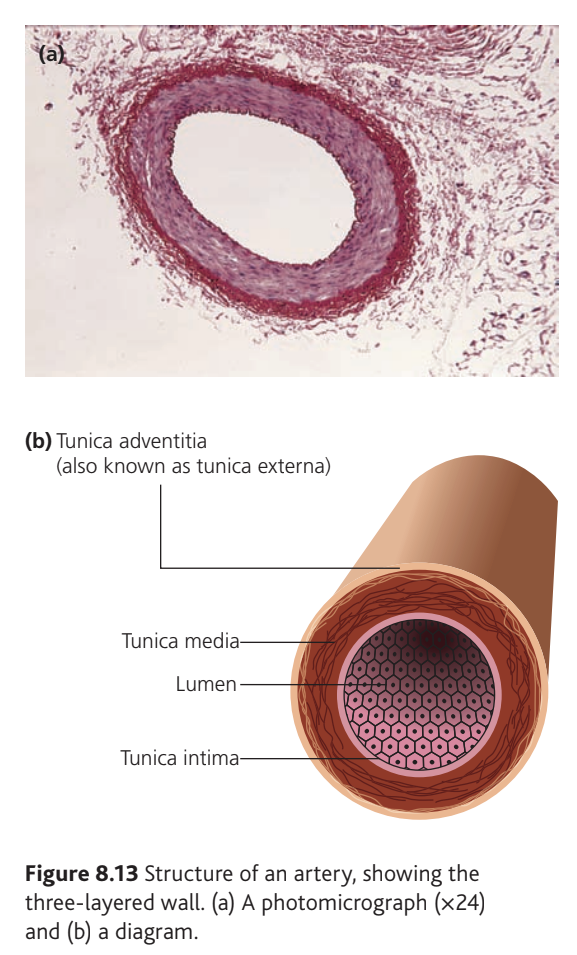
Three-Layered Artery Wall Structure
All arteries have a characteristic three-layered structure, each layer adapted to withstand high pressure and maintain blood flow. Understanding these layers is essential for comparing arteries with other vessel types.
Artery walls have a three-layered structure:
Tunica intima forms the innermost layer, lining the entire blood system. It consists of:
- An endothelium — a single layer of cells that lines the lumen (central cavity)
- A network of connective tissue
- A layer of elastic fibres
The endothelium's smooth surface reduces friction between the blood and vessel wall, allowing unrestricted blood flow.
Tunica media is the middle layer and is substantially thicker in arteries than in veins. This layer contains two key tissue types:
- Elastic tissue allows the blood vessel to stretch and recoil. This stretching slightly reduces pressure, while recoiling increases it again, which evens out pressure fluctuations and helps maintain blood pressure throughout the cardiac cycle.
- Smooth muscle tissue strengthens the artery wall to resist high blood pressure. The muscle can also contract to reduce blood flow. The body continuously redirects blood to areas of greatest need by controlling the contraction and relaxation of smooth muscle in arterioles.
Tunica adventitia (also called tunica externa) forms the outermost layer and consists mainly of the protein collagen. This tough material prevents the artery from over-stretching, which could damage the vessel wall.
Key Adaptations of Arteries
Each structural feature of arteries relates directly to function:
- Thick elastic tissue → stretches under pressure then recoils to maintain continuous flow
- Thick smooth muscle → resists high pressure and allows control of blood distribution
- Thick collagen layer → prevents vessel rupture from excessive stretching
Arterioles
As blood moves further from the heart, arteries branch repeatedly and become progressively smaller. The smallest arteries, leading directly into capillaries, are arterioles. These vessels play a vital role in regulating blood flow distribution.
Arteriole walls contain a high proportion of smooth muscle. When this muscle contracts, it narrows the vessel and restricts blood flow. The autonomic nervous system controls these muscle contractions, though the muscles also respond to local factors such as pH, oxygen levels, and carbon dioxide concentration.
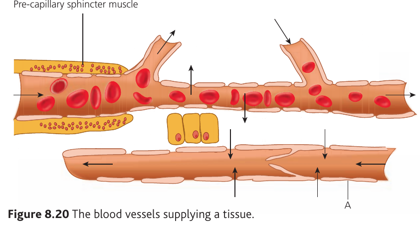
At the junction between arterioles and capillaries, rings of smooth muscle called pre-capillary sphincters regulate blood entry into capillary beds. Contraction of these sphincters can completely prevent blood from flowing into a particular capillary bed, allowing blood to bypass that area if necessary.
Blood Flow Regulation
Pre-capillary sphincters enable the body to redirect blood to areas of greatest need. For example, during exercise, sphincters open in muscle capillary beds while closing in the digestive system, ensuring active muscles receive maximum oxygen supply.
Capillaries
Capillaries serve as exchange sites where materials transfer between blood and cells. These vessels penetrate tissues extensively, reaching areas close to all cells. Capillaries are numerous and have a small cross-sectional area (small lumen).
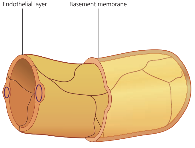
The small lumen creates friction with blood, which slows flow and lowers pressure. Both effects are important: slow flow provides more time for material exchange, while low pressure is necessary because capillary walls are extremely thin and weak to facilitate exchange.
Critical Capillary Adaptation
Capillary walls are only one cell thick, minimising the distance that materials must diffuse to enter or exit the vessel. This single-cell thickness is the key structural feature that enables efficient exchange of oxygen, nutrients, and waste products between blood and tissues.
White blood cells can squeeze through intercellular junctions, enabling them to enter tissues and combat infections.
Venules
Blood flows from capillaries into the smallest veins, called venules. The smallest venules consist only of an endothelium. Larger venules have a structure similar to veins but scaled down, with few or no elastic fibres.
Veins
Veins return blood to the heart. This blood is under low pressure, and the pulse present in arteries has disappeared. Veins have the same three layers as arteries, but the tunica media (containing muscle and elastic tissue) is much thinner.
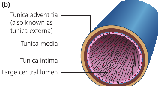
Several structural features relate to venous function:
Large lumen — This compensates for the absence of a pulse. Blood returning to the heart in veins must travel at the same rate as blood leaving in arteries, otherwise the heart would run out of blood. Although flow rate is slower in veins than arteries, the large lumen means the same volume of blood is delivered per unit time. The large lumen also reduces friction by ensuring much of the blood does not contact the vessel walls.
Thin tunica media — Low blood pressure means no thick muscular layer is needed to resist high pressure. Elastic tissue is reduced because vessels experience minimal stretching force (due to low pressure) and blood flow is even without pulsing.
Tunica adventitia — This layer is relatively thicker in veins than arteries because of the thinner tunica media. In actual thickness, however, it remains similar in both vessel types.
Valves — Veins contain valves along their length to ensure one-way flow towards the heart. Arteries do not require valves because the pulse pushes blood in the correct direction. When blood flows correctly, valve pockets flatten against the vein wall, allowing flow. If blood attempts to flow backwards, it fills the pockets, which bulge out and block the vein.
Thin walls — The force needed to push blood along veins is mainly produced when surrounding muscles contract during normal activity. Thin vein walls mean this external force is not resisted by the vessel itself.
How Veins Move Blood
Without a pulse to drive blood flow, veins rely on external forces. When skeletal muscles contract during movement, they squeeze veins and push blood toward the heart. Valves prevent backflow, ensuring blood only moves in the correct direction. This is why prolonged standing can cause blood pooling in the legs — without muscle contractions, venous return is reduced.
Summary of differences between arteries and veins
| Feature | Arteries | Veins |
|---|---|---|
| Direction of flow | Away from heart | Towards heart |
| Blood pressure | High | Low |
| Pulse | Present | Absent |
| Thickness of tunica media | Thick | Thin |
| Size of lumen | Narrow | Wide |
| Valves | Absent | Present |
Note that direction of flow, blood pressure and pulse presence are not structural differences but functional characteristics.
Blood, tissue fluid and lymph
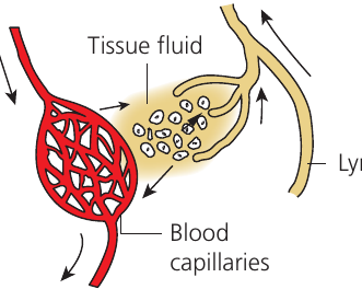
All substances required by cells must be in solution to be absorbed through membranes. Therefore, a liquid medium is needed at all stages of transport. Blood carries chemicals around the body, but transport between blood vessels and cells occurs via tissue fluid, which bathes and surrounds the cells. Excess tissue fluid is returned to the blood by a third fluid, lymph, which also plays a role in immunity.
Blood
Blood consists of the following components:
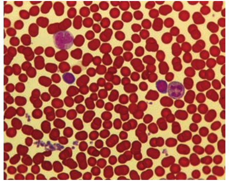
Red blood cells (erythrocytes) transport oxygen and play a role in carbon dioxide transport. These functions are carried out by the red pigment haemoglobin within the cells.
White blood cells (leukocytes) perform different roles in the immune system depending on their type.
Platelets are cell fragments important in the clotting process.
Plasma is the liquid medium of blood, which transports dissolved substances including amino acids, sugars, fatty acids, hormones and other proteins, clotting factors, and carbon dioxide.
Blood Component Functions
Each blood component has a specialized role:
- Red blood cells → oxygen and CO₂ transport (via haemoglobin)
- White blood cells → immune defense against pathogens
- Platelets → blood clotting to prevent blood loss
- Plasma → transport medium for dissolved nutrients, hormones, and waste products
Tissue fluid
Tissue fluid forms from plasma and contains many of the same solutes, with the important exception of larger protein molecules, which cannot escape from capillaries. Tissue fluid contains some white blood cells but no red blood cells or platelets.
Composition Difference
Tissue fluid is essentially plasma minus the large proteins. The capillary wall acts as a filter, allowing small dissolved molecules through but blocking larger protein molecules and blood cells (except some white blood cells which can actively squeeze through).
Lymph
Of the fluid leaving the blood to form tissue fluid, approximately is returned to the capillaries. The remaining returns to the bloodstream as lymph via the lymphatic system. Lymph is therefore very similar to tissue fluid but contains more white blood cells.
These white cells collect in large numbers in lymph nodes to fight infections, making them more common in lymph overall compared to tissue fluid.
Lymphatic system
The lymphatic system is a network of vessels and organs that connects to the circulatory system. Swellings called lymph nodes are found at intervals throughout the system. The tonsils, spleen and thymus gland are part of the lymphatic system.
As lymph flows through lymph nodes, some lymphocytes (B cells and T cells) and plasma cells are added to the fluid, along with antibodies produced by plasma cells. This explains the increased numbers of white cells and proteins found in lymph compared to tissue fluid.
Dual Role of Lymphatic System
The lymphatic system serves two important functions:
- Fluid balance → returns excess tissue fluid to the bloodstream
- Immunity → lymph nodes filter pathogens and produce antibodies, adding white blood cells and antibodies to the lymph
Comparison of blood, tissue fluid and lymph
| Component | Blood | Tissue fluid | Lymph |
|---|---|---|---|
| Red blood cells | Yes | No | No |
| White blood cells | Many | Few | Many |
| Proteins | Many | Few | More than tissue fluid (antibodies added) |
| Dissolved solutes | Yes | Yes | Yes |
Tissue fluid is formed from blood that has passed through gaps in capillary walls — effectively filtered. Larger molecules and structures therefore do not pass through. Blood cells do not pass through, except for some white blood cells which actively squeeze themselves through, particularly when tissue is damaged or infected.
Formation of tissue fluid
Capillaries are very leaky: approximately litres of fluid leaves the blood and enters the tissues every day. This fluid must be returned or the entire blood volume would be lost within an hour or two.
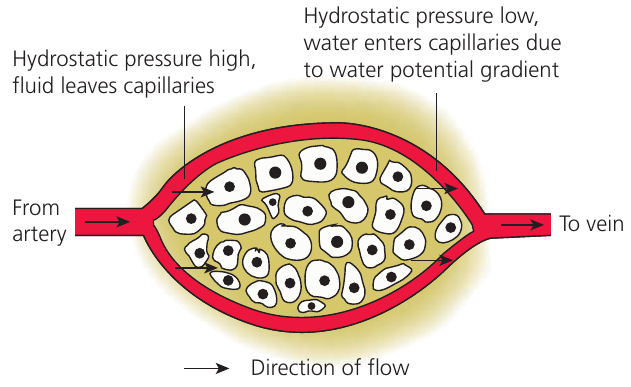
Two Opposing Forces Control Fluid Movement
Tissue fluid formation and recovery depend on the balance between two forces:
- Hydrostatic pressure (blood pressure) → forces fluid out of capillaries
- Oncotic pressure gradient (water potential) → draws water into capillaries by osmosis
The balance between these forces changes along the length of the capillary, determining whether fluid leaves or enters the blood.
Two factors control fluid movement to and from blood capillaries:
Hydrostatic pressure of the blood (i.e. blood pressure) tends to force fluid out.
Oncotic pressure gradient (related to water potential) tends to draw water in by osmosis. The water potential of blood is lower than that of tissue fluid, mainly because of plasma proteins, most of which do not enter tissue fluid.
Blood hydrostatic pressure drops considerably as blood passes through capillaries:
At the arterial end of the capillary bed, hydrostatic pressure is relatively high. This overcomes the water potential gradient, forcing fluid through the capillary walls. No blood cells or large protein molecules leave because they are too large to pass through gaps in the capillary walls.
At the venous end of the capillary bed, the water potential gradient remains largely unchanged, but the drop in hydrostatic pressure means it is now less than the pressure due to the water potential gradient. As a result, water re-enters the blood.
Fluid Movement Mechanism
- Arterial end: High hydrostatic pressure > water potential gradient → fluid forced out
- Venous end: Low hydrostatic pressure < water potential gradient → water drawn back in
- Net result: Approximately 90% of fluid is recovered; remaining 10% returns via lymphatic system
Approximately of fluid lost at the arterial end is recovered at the venous end. The remaining is collected by lymph vessels, which eventually return it to the blood.
Key terms
Hydrostatic pressure is the pressure exerted by a fluid, for example the blood.
Oncotic pressure is a form of osmotic pressure exerted by proteins, notably albumin, in a blood vessel's plasma, that usually tends to pull water into the circulatory system.
Key Points to Remember:
-
Blood vessels have structures adapted to their specific functions: arteries (high pressure transport), capillaries (exchange), and veins (low pressure return)
-
The three vessel wall layers are tunica intima (endothelium), tunica media (muscle and elastic tissue), and tunica adventitia (collagen)
-
Tissue fluid forms when hydrostatic pressure forces plasma out of capillaries at the arterial end; water potential gradient draws water back at the venous end
-
Blood contains all cell types (RBCs, WBCs, platelets) and proteins; tissue fluid lacks large proteins and most cells; lymph contains many WBCs and some proteins
-
Pre-capillary sphincters regulate blood flow into capillary beds, allowing the body to direct blood where most needed
-
The one-cell-thick capillary wall is the critical structural feature enabling efficient material exchange between blood and tissues
-
Approximately 20 litres of fluid leave the blood daily, with 90% recovered at the venous end and 10% returned via the lymphatic system