Cell Division, Diversity, and Organisation (OCR A-Level Biology A): Revision Notes
The Cell Cycle
Introduction to the cell cycle
Following cell division, newly formed cells require time to mature before they can divide again. This repeating pattern of division, growth, maturation, and subsequent division is known as the cell cycle. The duration of this cycle varies significantly between different cell types, with more specialized cells typically exhibiting longer cycles.
Certain highly specialized cells, including muscle cells and nerve cells, lose their capacity to divide entirely. Mammalian red blood cells also cannot divide, as they lose their nucleus during their development process. This loss of mitotic capability reflects the trade-off between specialization and the ability to reproduce.
Duration of the cell cycle across cell types
The length of the cell cycle differs considerably depending on the cell type. Less specialized cells, such as embryonic cells, divide rapidly with cycle times as short as 8-60 minutes. In contrast, highly differentiated cells like hepatocytes (liver cells) may have cycles lasting approximately one year. This variation reflects the different functional demands placed on various tissues within an organism.
| Cell type | Duration of cell cycle |
|---|---|
| Embryo cells | 8–60 minutes |
| Yeast cells | 1.5–3 hours |
| Intestinal epithelial cells | about 12 hours |
| Bone marrow cells | about 18 hours |
| Stomach epithelial cells | about 24 hours |
| Hepatocytes (liver cells) | about 1 year |
The table demonstrates that rapidly dividing tissues, such as those in the intestinal lining or bone marrow, have much shorter cell cycles than slower-renewing tissues. This reflects the body's need to constantly replace cells in high-turnover areas while maintaining stability in more permanent tissues.
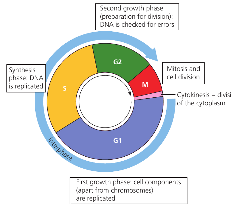
Phases of the cell cycle
The cell cycle comprises several distinct phases, each with specific functions in preparing the cell for division and ensuring accurate replication of genetic material.
Interphase
Interphase represents the portion of the cell cycle when the cell is not actively dividing. This extended period encompasses three sub-phases and constitutes approximately 95% of the total cell cycle duration. During interphase, the cell grows, carries out its normal metabolic functions, and prepares for division.
G₁ phase (first growth phase)
Following cell division, the newly formed cell enters the G₁ phase (first growth phase). During this stage, the cell synthesizes proteins, produces organelles, and increases in size. Essentially, all cellular components except the chromosomes are replicated. The cell accumulates the materials and energy required for DNA synthesis and eventual division.
This phase is highly variable in length and accounts for much of the difference in cell cycle duration between different cell types.
S phase (synthesis phase)
The S phase (synthesis phase) is characterized by DNA replication. Each chromosome is duplicated through semi-conservative replication, producing two identical sister chromatids joined at the centromere. This ensures that when the cell divides, each daughter cell receives a complete set of genetic information. The duration of S phase is relatively constant across different cell types, typically lasting several hours.
G₂ phase (second growth phase)
After DNA replication, the cell enters the G₂ phase (second growth phase). This represents the final preparation stage before mitosis. During G₂, the cell continues to grow and produces proteins necessary for cell division, particularly those involved in chromosome condensation and spindle formation.
Enzymes check the newly replicated DNA for errors that may have occurred during replication. At the conclusion of G₂, the DNA begins to condense tightly, becoming visible as distinct chromosomes as mitosis commences.
M phase (mitosis)
The M phase encompasses mitosis—the division of the nucleus—and represents only approximately 5% of the cell cycle, though this proportion varies between cell types. During mitosis, the replicated chromosomes are separated and distributed equally to two daughter nuclei. This process is divided into several stages (prophase, metaphase, anaphase, and telophase), each with characteristic chromosome movements and cellular changes.
Cytokinesis
Cytokinesis refers to the division of the cytoplasm following nuclear division. This process physically separates the parent cell into two daughter cells, each receiving approximately half of the cytoplasm and organelles.
In animal cells, cytokinesis occurs through the formation of a cleavage furrow that pinches the cell in two. In plant cells, a cell plate forms between the two nuclei, eventually developing into a new cell wall.
Cell cycle checkpoints
The cell cycle incorporates quality control mechanisms at specific points called checkpoints. These checkpoints monitor the integrity of genetic information and ensure proper progression through the cycle. If errors are detected, the cycle may be paused to allow for repair, or the cell may undergo programmed cell death (apoptosis) to prevent transmission of mutations.
G₁ checkpoint
During the G₁ checkpoint, chromosomes are examined for damage. If damage is detected, the cell does not progress into S phase until the DNA has been successfully repaired. This prevents replication of damaged genetic material.
S phase checkpoint
A verification process during the S phase checkpoint confirms that all chromosomes have been completely replicated. If replication is incomplete, the cell cycle is halted. This ensures that both daughter cells will receive a full complement of genetic information.
G₂ checkpoint
The G₂ checkpoint conducts another examination for DNA damage that may have occurred during the replication process. The cycle may be delayed at this stage to allow for DNA repair, preventing the transmission of replication errors to daughter cells.
Metaphase checkpoint
The final metaphase checkpoint occurs during mitosis itself. This checkpoint verifies that all chromosomes have correctly attached to the spindle fibres before allowing progression to anaphase. This prevents chromosome loss or unequal distribution during cell division.
These checkpoints are mediated by specialized enzymes with proof-reading and repair functions. They represent a critical safeguard against the development of mutations and cellular abnormalities. When checkpoint mechanisms fail, uncontrolled cell division can occur, potentially leading to conditions such as cancer.
DNA levels during the cell cycle
The quantity of DNA within a dividing cell changes in a predictable pattern throughout the cell cycle. This can be measured using flow cytometry, a technique that employs fluorescent dyes binding to nucleic acids. The fluorescence intensity correlates with DNA content—greater fluorescence indicates more DNA present.
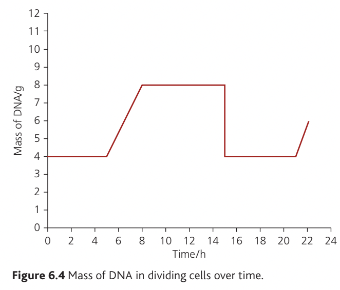
DNA Content Changes During the Cell Cycle:
The graph demonstrates DNA content changes over a 24-hour period in dividing cells:
- G₁ phase (hours 0-4): DNA content remains constant at baseline level (~4 µg)
- S phase (hours 4-8): DNA content doubles as replication occurs, reaching ~8 µg
- G₂ phase (hours 8-16): DNA content remains at elevated level (8 µg) while cell prepares for division
- M phase (hour 16): DNA content returns to baseline (4 µg) as genetic material is distributed equally between two daughter cells
- The pattern then repeats as new cells enter their own cell cycles
This predictable pattern of DNA doubling and halving confirms that mitosis produces two daughter cells with identical genetic content to the parent cell. The consistent return to baseline DNA levels distinguishes mitosis from meiosis, where DNA content would be reduced by half in the gametes produced.
Key Points to Remember:
- The cell cycle is the sequence of events from one cell division to the next, varying from minutes to years depending on cell type
- Interphase (G₁, S, and G₂ phases) occupies approximately 95% of the cell cycle, while mitosis takes only about 5%
- S phase is when DNA replication occurs, doubling the cell's genetic content
- Four checkpoints (at G₁, S, G₂, and metaphase) monitor DNA integrity and ensure accurate cell division
- Some specialized cells (muscle, nerve, red blood cells) lose the ability to divide during their development