Homologous Chromosomes and Meiosis (OCR A-Level Biology A): Revision Notes
Homologous Chromosomes and Meiosis
What are homologous chromosomes?
A complete set of chromosomes consists of multiple homologous pairs. In humans, there are chromosomes arranged into homologous pairs.
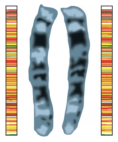
A homologous pair refers to two chromosomes that carry the same sequence of genes. One chromosome in each pair originates from the mother (maternal chromosome), while the other comes from the father (paternal chromosome).
Each homologous pair contains chromosomes of the same length, centromere position, and gene sequence, but they may carry different versions (alleles) of those genes. This pairing is fundamental to sexual reproduction and genetic diversity.
Why are homologous pairs important?
During sexual reproduction, gametes (sex cells) fuse to form a zygote. Each gamete must contain only one set of chromosomes—the haploid number (n). When two gametes combine, the zygote receives the full diploid number (2n), matching the chromosome count of the parent organisms.
Homologous chromosomes ensure that every gene is represented in the gamete. Although both chromosomes in a pair carry the same genes, they may contain different versions called alleles. For example, a person might have one chromosome carrying a 'blue' eye colour allele and its homologous partner carrying a 'brown' allele.
Visualising homologous chromosomes
Homologous chromosomes have identical structures and staining properties, making it possible to identify and pair them from microscope images. A karyogram displays all chromosomes arranged in homologous pairs.
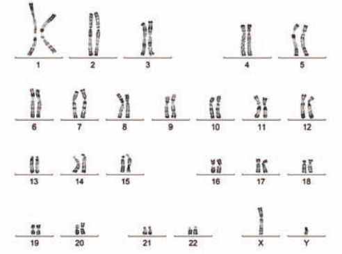
The exception to matching structures occurs with sex chromosomes in males, where the X and Y chromosomes differ significantly in size and gene content. Despite these differences, they still function as a homologous pair during meiosis.
Stages of meiosis
Meiosis involves two successive divisions that produce four haploid cells from one diploid cell. The first division (Meiosis I) reduces the chromosome number by half, while the second division (Meiosis II) resembles mitosis.
Overview of meiosis stages
| Stage | Key events |
|---|---|
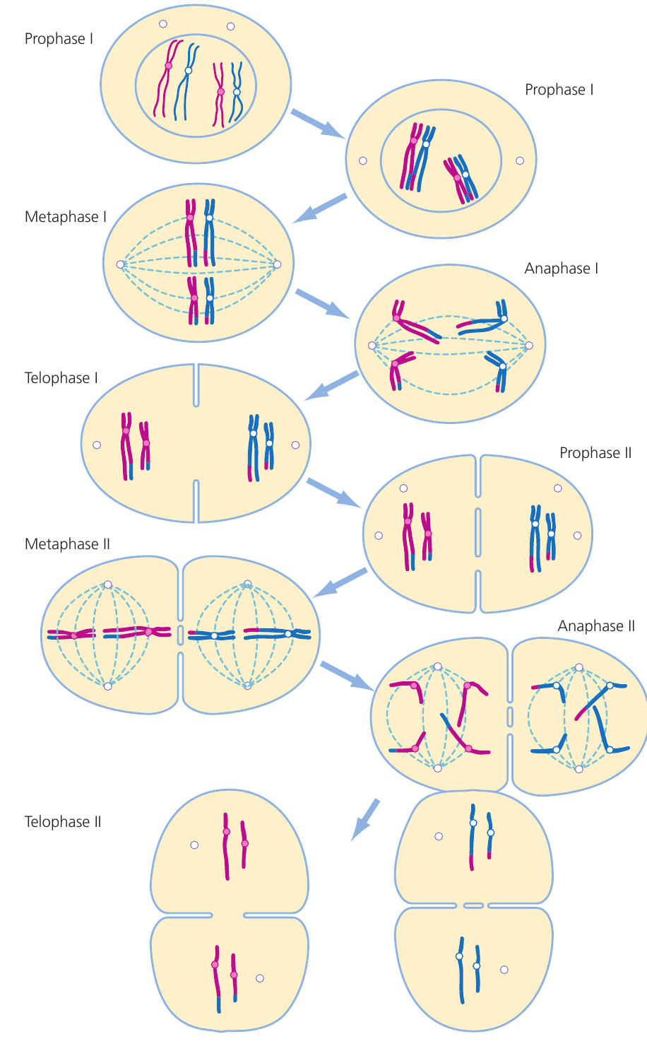
| Prophase I | Chromosomes condense as sister chromatids. Homologous chromosomes pair up to form tetrads. Crossing over may occur between non-sister chromatids. Centrioles migrate to opposite poles. |
| Metaphase I | Tetrads align at the spindle equator. |
| Anaphase I | Homologous chromosomes separate and move to opposite poles. Sister chromatids remain joined. |
| Telophase I | Each pole receives a haploid number of chromosomes. Nuclear membranes may reform. |
| Cytokinesis I | Cytoplasm divides, forming two haploid cells. No DNA replication occurs. |
| Prophase II | Nuclear membrane breaks down (if reformed). |
| Metaphase II | Individual chromosomes (each consisting of two sister chromatids) align at the spindle equator in each cell. |
| Anaphase II | Sister chromatids separate and move to opposite poles. |
| Telophase II | Chromosomes decondense and nuclear membranes reform. |
| Cytokinesis II | Cytoplasm divides, creating four genetically different haploid cells. |
Detailed description of meiosis I
Prophase I begins with chromosome condensation, making them visible under light microscopy. Homologous chromosomes then pair up to form structures called bivalents or tetrads (containing four chromatids). Points of contact called chiasmata (singular: chiasma) form where non-sister chromatids can exchange genetic material through crossing over. The nuclear membrane fragments and becomes incorporated into the endoplasmic reticulum, while centrioles replicate and migrate to opposite poles, forming spindle microtubules.
The formation of bivalents during Prophase I is unique to meiosis and does not occur in mitosis. This pairing allows homologous chromosomes to align precisely for separation and creates opportunities for crossing over.
During Metaphase I, bivalents position themselves randomly at the cell's equatorial plate. Microtubules from the spindle apparatus attach to the centromere of each chromosome. The independent positioning of maternal and paternal chromosomes is crucial for genetic variation.
In Anaphase I, entire chromosomes (each still consisting of two sister chromatids) are pulled toward opposite poles by shortening microtubules. This differs from mitosis, where sister chromatids separate during anaphase.
During Anaphase I, whole chromosomes (consisting of two sister chromatids joined at the centromere) move to opposite poles. Sister chromatids do NOT separate in Meiosis I—they remain attached and only separate during Meiosis II. This is a critical difference from mitosis.
Telophase I sees chromosomes arriving at opposite poles. Nuclear membranes reform around two daughter nuclei, each containing the haploid number of chromosomes. Cytokinesis occurs as the cell membrane constricts, creating two separate cells with small cytoplasmic bridges between them.
An interphase may occur between the two meiotic divisions, during which chromosomes may uncoil. However, no DNA replication takes place between Meiosis I and II.
Detailed description of meiosis II
The second meiotic division closely resembles mitosis but occurs in haploid cells.
Prophase II involves centriole replication and migration to poles positioned at right angles to those in Meiosis I. The nuclear membrane breaks down again.
During Metaphase II, individual chromosomes align at the equator. If crossing over occurred during Meiosis I, chromatids arrange themselves randomly—this is particularly important for generating variation.
In Anaphase II, sister chromatids finally separate at the centromere and migrate to opposite poles, just as they would in mitosis.
Telophase II sees nuclear membranes reforming and cells dividing through cytokinesis, ultimately producing four haploid cells that are genetically distinct from each other and from the parent cell.
Key differences between meiosis and mitosis
Meiosis I shows two critical differences from mitosis:
- Homologous chromosomes pair up during Prophase I and separate into different cells during Anaphase I. This pairing does not occur in mitosis.
- Sister chromatids remain attached throughout Meiosis I and only separate during Meiosis II. In mitosis, sister chromatids separate during anaphase.
The second division of meiosis closely resembles mitosis because sister chromatids separate, maintaining chromosome number (though the cells are haploid). This is why Meiosis II is sometimes described as "mitosis in haploid cells."
Independent assortment, crossing over and genetic variation
Sexual reproduction through meiosis produces gametes showing considerable genetic variation, which provides important evolutionary advantages.
Independent assortment
During Meiosis I, one chromosome from each homologous pair enters each gamete. Independent assortment describes the random nature of this process—which chromosome from any pair ends up in a particular gamete is entirely random.
Consider a parent cell with two homologous pairs (four chromosomes total). Different combinations of maternal and paternal chromosomes can be distributed to gametes. The number of possible combinations increases dramatically with more chromosome pairs.
Worked Example: Calculating Genetic Combinations from Independent Assortment
In humans, with pairs of chromosomes, independent assortment alone generates over million different possible combinations.
Step 1: Identify the formula Number of combinations = where is the number of chromosome pairs
Step 2: Substitute values Number of combinations =
Step 3: Calculate the result possible combinations
This means independent assortment alone can produce over 8 million genetically different gametes from a single individual!
When fertilisation occurs, not only is the diploid number restored, but homologous pairs are also re-established. Since homologous chromosomes carry the same genes but potentially different alleles, each gamete—and therefore each offspring—has a unique genetic composition.
Crossing over
During Prophase I, crossing over adds another layer of genetic variation. Non-sister chromatids from homologous chromosomes exchange segments of genetic material at chiasmata. This creates new combinations of alleles on individual chromosomes that did not exist in either parent.
Crossing over occurs between non-sister chromatids (chromatids from different homologous chromosomes), NOT between sister chromatids. This ensures that genetic material is exchanged between maternal and paternal chromosomes, creating novel allele combinations.
Sources of variation in meiosis
Meiosis generates genetic variation through:
- Independent assortment: Random distribution of maternal and paternal chromosomes into gametes
- Crossing over: Exchange of genetic material between non-sister chromatids during Prophase I
- Random fertilisation: Any gamete can potentially fuse with any other during fertilisation
These mechanisms ensure that offspring are genetically unique, providing raw material for evolution and adaptation.
Key Points to Remember:
- Homologous chromosomes carry the same genes in the same sequence but may have different alleles. One comes from each parent.
- Meiosis involves two divisions: Meiosis I separates homologous pairs (reduction division), while Meiosis II separates sister chromatids (similar to mitosis).
- The first division is unique to meiosis—homologous chromosomes pair up during Prophase I and separate during Anaphase I, while sister chromatids remain together.
- Independent assortment and crossing over during Meiosis I create genetic variation, producing genetically unique haploid gametes.
- Meiosis produces four genetically different haploid cells from one diploid parent cell, essential for sexual reproduction.