The Microscope (Leaving Cert Biology): Revision Notes
📚 Revision Notes
The Microscope
- The basic unit of structure is the cell.
- Cells are often described as the building blocks of life.
- Every cell arises from pre-existing cells.
- Cells are tiny structures measured in micrometres (µm). They can only be viewed under a microscope.
infoNote
Did you know? In 1665, Robert Hooke, while studying cork under a microscope, saw tiny box-like structures. These reminded him of the rooms where monks lived, called "cellula." He coined the term "cell" to describe what he saw, marking the first use of the word in a scientific context.
1. The Compound Light Microscope (can only view the nucleus)
2. The Electron Microscope (to view cell ultrastructures)
1. The Compound Light Microscope
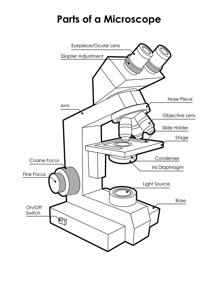
| Eyepiece Lens | Magnifies the object. |
|---|---|
| Coarse Focus Wheel | For rough focusing. |
| Fine Focus Wheel | Brings the specimen into clear focus. |
| Stage Clips | To hold the slide. |
| Nosepiece | Revolves to move the desired lens into place. |
| Objective Lens | Magnifies the image. |
| Stage | Holds the slide. |
| Diaphragm | Controls the amount of light passing through the specimen. |
| Mirror/Light Source | Reflect light through the stage, allowing the specimen to be seen more clearly. |
Magnification
To calculate the total magnification, multiply the magnification of the eyepiece lens by the magnification of the objective lens.
| Eyepiece lens | Objective lens | Total magnification |
|---|---|---|
| x10 | x4 | x40 |
| x15 | x10 | x150 |
2. Electron Microscope
An electron microscope allows the cell ultrastructure to be seen (the cell organelle).
- Transmission Electron Microscope (TEM): Sends a beam of electrons through a thin specimen to reveal the internal structure in great detail.
- Scanning Electron Microscope: Sends a beam of electrons over the surface of the specimen to view its outer structure.