The Human Eye (Grade 12 NSC Matric Life Sciences): Revision Notes
The Human Eye
Introduction to the human eye
The human eye is one of our most important sense organs, containing a high concentration of receptor cells that can detect light stimuli and produce responses. As humans, we have two eyes positioned at the front of our skull that work together to provide us with clear vision and help us navigate our environment effectively.
Key terminology
Understanding the basic terminology of the eye is essential for studying how this complex organ functions. Let's start with the fundamental terms you need to know.
The following key terms form the foundation of understanding how the human eye works. Pay special attention to the differences between rods and cones, as these are frequently tested concepts.
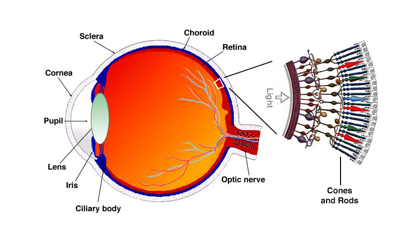
The eye contains two main types of light-detecting cells called photoreceptors. Rods are specialised receptor cells located in the retina that are highly sensitive to dim light conditions. These cells help us see in low-light environments and are responsible for our black and white vision. Cones, on the other hand, are receptor cells that work best in bright light conditions and enable us to distinguish between different colours, giving us our colour vision.
The pupil serves as the central opening within the iris that controls how much light enters the eye. Think of it as the gateway that allows light to pass through to the internal structures of the eye.
Several important mechanisms control how our eyes function. The pupillary mechanism refers to how the pupil size changes to regulate the amount of light entering the eye - much like how a camera aperture opens and closes. Accommodation is the remarkable ability of the eye's lens to change its shape, allowing us to focus clearly on objects whether they are near to us or far away.
The field of vision describes the total area that one eye can see at any given moment. When we talk about lens shapes, convex refers to a shape that curves outward and is thicker in the middle than at the edges, while concave describes a shape that curves inward and is thinner in the middle than at the edges.
Structure of the eye
The human eye has a sophisticated structure designed to capture light, focus it precisely, and convert it into electrical signals that our brain can interpret as vision.
The eye's structure can be thought of like a sophisticated camera system, with each component playing a specific role in creating the images we see.
The eye consists of several layers and components working together. The outer protective structures include the eyelids and eyelashes, which help protect the eye from foreign particles and mechanical injury. The white outer covering you can see is called the sclera, while the coloured part of the eye is the iris, with the pupil at its centre.
Internally, the eye can be divided into three main layers: the sclera (outer layer), the choroid (middle layer), and the retina (inner layer). Each layer has specific adaptations that make vision possible.
Functions of eye structures
Each part of the eye has evolved specific structural features that enable it to perform particular functions in the vision process.
The sclera forms the tough, white, inelastic outer layer that covers most of the eyeball. Its strong, inflexible nature provides excellent protection for the delicate internal structures while maintaining the eye's spherical shape, which is crucial for proper vision.
The cornea is the transparent front portion of the sclera that has a distinctive convex (bulging outward) shape. This transparency allows light to pass through easily, while its curved shape causes the incoming light rays to bend (refract), beginning the focusing process that will eventually create a clear image on the retina.
The cornea performs the majority of the eye's focusing power due to its curved shape. Any damage or irregularity to the cornea can significantly affect vision quality.
The choroid is a dark, pigmented middle layer containing numerous blood vessels. The dark pigmentation prevents light from bouncing around inside the eye, which would cause blurred vision, while the blood vessels provide essential nutrients and oxygen to the retinal cells.
The ciliary body contains ciliary muscles and forms a thickened region at the front part of the choroid. These muscles can contract or relax to change the tension on the suspensory ligaments, which is crucial for the accommodation process.
Suspensory ligaments connect the ciliary body to the lens. When the tension in these ligaments changes, it alters the shape of the lens, allowing the eye to focus on objects at different distances.
The iris is the coloured portion of the eye with the pupil at its centre. It contains two types of muscles - circular and radial - that work together to control the size of the pupil and regulate how much light enters the eye.
The iris colour is determined by the amount and type of pigment present. Brown eyes have more pigment, while blue eyes have less pigment and scatter light differently.
The retina forms the inner light-sensitive layer of the eye and contains both rods and cones. Rods respond to low-intensity light and provide night vision as well as peripheral vision, while cones respond to bright light and provide sharp, clear colour vision. Neurons in the retina carry impulses from the rods and cones through the optic nerve to the brain.
The yellow spot is a small area on the retina containing the highest concentration of cone cells. This region provides us with our clearest, sharpest vision and is what we use when we need to see fine details.
The blind spot is a small area on the retina where the optic nerve exits the eye. This region contains no rods or cones, creating a small area where we cannot detect light. However, our brain compensates for this, and we normally don't notice this gap in our vision.
The lens is an elastic, transparent, biconvex structure positioned behind the pupil. Its ability to change shape allows the eye to focus on both near and distant objects, and its transparency ensures light can pass through without obstruction.
Two types of fluid help maintain the eye's shape and assist in light refraction. Aqueous humour is the watery fluid found between the cornea and lens that maintains the cornea's shape and plays a small role in bending light. Vitreous humour is a jelly-like substance found behind the lens that maintains the eyeball's shape and also contributes slightly to light refraction.
Accommodation mechanism
Accommodation is one of the most remarkable features of human vision - our ability to automatically adjust our focus when looking at objects at different distances.
Worked Example: How Accommodation Works
For near objects (less than 6 metres):
- Step 1: Ciliary muscles contract
- Step 2: Suspensory ligaments become slack (loose)
- Step 3: Lens becomes more convex (rounded)
- Step 4: Light rays bend more sharply to focus on retina
For distant objects (more than 6 metres):
- Step 1: Ciliary muscles relax
- Step 2: Suspensory ligaments become taut (tight)
- Step 3: Lens becomes flatter and less convex
- Step 4: Light rays bend less to focus on retina
The accommodation process works differently depending on whether we're viewing near or distant objects. When we look at something close to us (less than 6 metres away), our ciliary muscles contract. This contraction causes the suspensory ligaments to become slack (loose), which reduces the tension on the lens. As a result, the lens becomes more convex (more rounded), which bends the incoming light rays more sharply to focus them properly on the retina.
When viewing distant objects (more than 6 metres away), the opposite happens. The ciliary muscles relax, causing the suspensory ligaments to become taut (tight). This increases the tension on the lens, making it flatter and less convex. The flatter lens bends light rays less, which is exactly what's needed to focus distant objects clearly on the retina.
This process happens automatically and very quickly, allowing us to shift our focus from a book in our hands to a bird flying in the distance without conscious effort.
Pupillary mechanism
The pupillary mechanism is our eye's automatic system for controlling how much light enters, ensuring we can see clearly in both bright and dim conditions.
Worked Example: Pupillary Response
In bright light:
- Circular muscles of iris: Contract
- Radial muscles of iris: Relax
- Pupil response: Constricts (becomes smaller)
- Result: Less light enters, protecting retina
In dim light:
- Circular muscles of iris: Relax
- Radial muscles of iris: Contract
- Pupil response: Dilates (becomes larger)
- Result: More light enters, improving vision
The iris contains two types of muscles that work as antagonistic pairs - when one contracts, the other relaxes. In bright light conditions, the circular muscles of the iris contract while the radial muscles relax. This causes the pupil to constrict (become smaller), reducing the amount of light entering the eye and preventing damage to the sensitive retina.
In dim light conditions, the opposite occurs. The radial muscles contract while the circular muscles relax, causing the pupil to dilate (become larger). This allows more light to enter the eye, improving our ability to see in low-light conditions.
This mechanism is controlled by the autonomic nervous system and happens involuntarily - you can observe it by looking in a mirror and turning a light on and off, watching how your pupils respond.
Types of vision
Having two eyes provides us with significant advantages over single-eye vision, giving us enhanced depth perception and a wider field of view.
Binocular vision occurs because we have two eyes with overlapping fields of vision. Each eye sees a slightly different image of the same object, and our brain combines these separate images and interprets them as one complete image. This overlap is crucial for proper vision.
Stereoscopic vision refers to our ability to form three-dimensional images from the slightly different perspectives provided by each eye. This gives us the ability to judge distance, depth, and the relative size of objects accurately. This is why people with vision in only one eye may have difficulty with tasks requiring depth perception, such as catching a ball or judging distances while driving.
Visual defects
While the human eye is remarkably sophisticated, various defects can affect vision quality.
Myopia (Short-sightedness)
Myopia occurs when a person can see nearby objects clearly but struggles with distant objects. This happens because light rays from distant objects focus in front of the retina rather than directly on it, causing blurred vision.
Common causes:
- An eyeball that is too long
- A cornea that is too curved for the eyeball's length
- A lens that cannot become flat enough
Treatment: Wearing glasses with concave lenses to help focus light correctly on the retina.
Myopia (short-sightedness) occurs when a person can see nearby objects clearly but struggles with distant objects. In this condition, light rays from distant objects focus in front of the retina rather than directly on it, causing blurred vision. This can be caused by an eyeball that is too long, a cornea that is too curved for the eyeball's length, or a lens that cannot become flat enough. Treatment typically involves wearing glasses with concave lenses to help focus light correctly on the retina.
Key Points to Remember:
-
Rods work in dim light for black and white vision, while cones work in bright light for colour vision - this is why we can't see colours well in very dim lighting.
-
Accommodation involves the lens changing shape - more convex for near objects, less convex for distant objects, controlled by ciliary muscles and suspensory ligaments working together.
-
The pupillary mechanism automatically adjusts pupil size - smaller in bright light to protect the retina, larger in dim light to gather more light.
-
The retina contains all our light-sensitive cells, with the yellow spot providing our sharpest vision and the blind spot creating a small area where we cannot see.
-
Binocular vision gives us depth perception - two eyes working together provide better distance judgement and a wider field of view than single-eye vision.