Nuclear Magnetic Resonance Spectroscopy (VCE SSCE Chemistry): Revision Notes
Nuclear Magnetic Resonance Spectroscopy
Nuclear Magnetic Resonance (NMR) spectroscopy is one of the most powerful techniques for determining the structure of complex molecules. When combined with infrared (IR) spectroscopy and other analytical methods, NMR can help you work out the exact structure of an unknown organic compound.
What is NMR spectroscopy?
NMR spectroscopy uses electromagnetic radiation in the radio frequency range to gather information about molecular structure. Unlike other forms of spectroscopy you've studied, the energy of radio waves is too low to cause electronic, vibrational, or rotational transitions in molecules. Instead, NMR detects changes in nuclear spin states.
Medical application: MRI
A form of NMR technology called Magnetic Resonance Imaging (MRI) is used in medicine to create highly detailed pictures of internal body structures. MRI can detect diseased tissue without using harmful radiation like X-rays.
Principles of nuclear magnetic resonance spectroscopy
Nuclear spin
For atoms to be detected by NMR spectroscopy, their nuclei must have a property called nuclear spin. Only nuclei with an odd number of protons and/or neutrons possess nuclear spin. The most important examples for organic chemistry are:
- Hydrogen-1 (H) - one proton, no neutrons
- Carbon-13 (C) - six protons, seven neutrons
- Phosphorus-31 (P) - fifteen protons, sixteen neutrons
The odd number of nuclear particles causes these nuclei to behave like tiny bar magnets.
Energy transitions in a magnetic field
When placed in an external magnetic field, nuclei with spin can adopt one of two orientations:
- Lower energy state: aligned with the magnetic field
- Higher energy state: aligned against the magnetic field
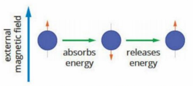
Inside an NMR spectrometer, nuclei normally exist in the lower energy state. A radio frequency transmitter provides energy to 'flip' nuclei into the higher energy state. Over time, the nuclei naturally return to the lower energy state, releasing a pulse of energy as they do so. This released energy is detected and presented as an NMR spectrum.
The energy difference between the two spin states depends on:
- The type of nucleus being examined
- The chemical environment surrounding the nucleus
This dependence on chemical environment is what makes NMR such a powerful tool for structure determination!
Types of NMR spectroscopy
Proton (H) NMR spectroscopy is the most common form. It examines hydrogen-1 nuclei and provides information about any molecule containing hydrogen atoms. Note that chemists often use the terms 'hydrogen' and 'proton' interchangeably when discussing NMR.
Carbon-13 (C) NMR spectroscopy examines carbon-13 nuclei and is valuable for investigating the carbon backbone of organic molecules.
Chemical environments
What is a chemical environment?
A chemical environment is made up of the atoms and electrons that surround a specific atom in a molecule. Atoms are in the same chemical environment if they are:
- Attached in the same way
- Bonded to the same types of atoms
- Positioned symmetrically within the molecule
Atoms in the same chemical environment are called equivalent atoms. All equivalent atoms produce a single signal in an NMR spectrum.
Key Principle: The number of signals in an NMR spectrum equals the number of different chemical environments.
Examples of chemical environments
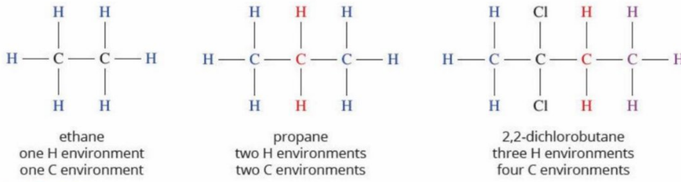
Worked Example: Identifying Chemical Environments
Ethane ():
- All six hydrogen atoms are equivalent (each is part of a group)
- The two carbon atoms are equivalent
- Result: ONE hydrogen signal and ONE carbon signal
Propane ():
- Six hydrogen atoms in the groups are equivalent
- Two hydrogen atoms in the central group form a different environment
- Result: TWO hydrogen signals and TWO carbon signals
2,2-dichlorobutane:
- Three different hydrogen environments
- Four different carbon environments
- Result: THREE hydrogen signals and FOUR carbon signals
General rule: Molecules with more symmetry have fewer different chemical environments. The more planes of symmetry a molecule has, the fewer unique chemical environments it contains.
How NMR spectrometers work
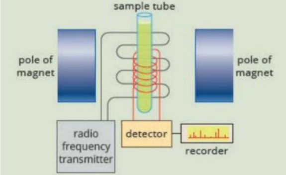
An NMR spectrometer contains:
- A powerful magnet that creates a strong external magnetic field
- A spinning sample tube (ensures uniform magnetic field exposure)
- A radio frequency transmitter (provides energy to flip nuclear spins)
- A detector (measures energy released as nuclei return to lower energy states)
- A computer (analyses signals and produces the NMR spectrum)
The sample is dissolved in a special solvent that doesn't produce NMR signals. Common solvents include , , and . These contain deuterium (H), oxygen-16, or carbon-12, which don't show up in proton or carbon-13 NMR spectroscopy.
Interpreting proton NMR spectra
A proton NMR spectrum provides four key pieces of information:
- Number of signals - shows how many different hydrogen environments exist
- Relative peak area - indicates the number of hydrogen atoms in each environment
- Signal splitting pattern - reveals information about neighbouring hydrogen atoms
- Chemical shift - helps identify the type of chemical environment
Peak area and integration
The area under each signal (called the peak area) is proportional to the number of hydrogen atoms in that environment. The ratio of peak areas tells you the ratio of hydrogen atoms in each environment.
You may need to multiply the ratio by a factor to account for all hydrogen atoms in the molecule.
Worked Example: 2,2,4,4-tetrachloropentane
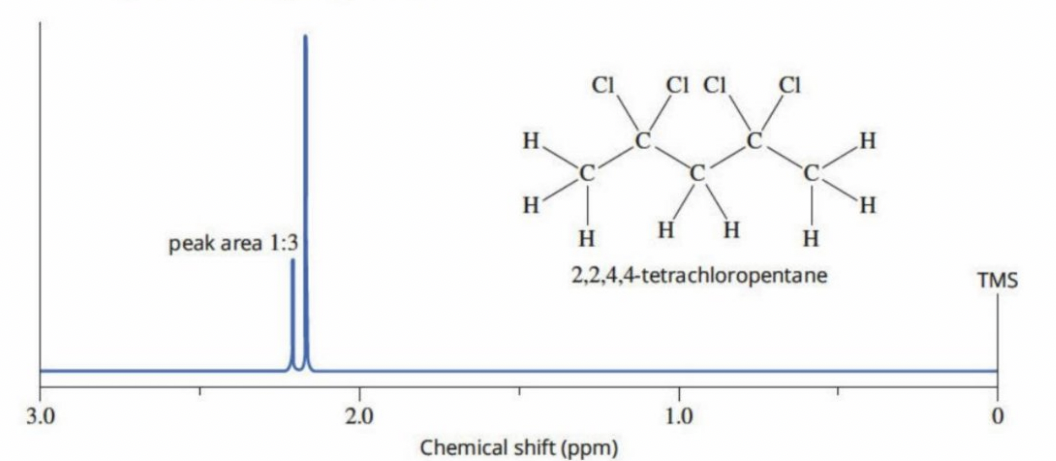
This molecule shows two signals with relative peak areas of 1:3.
- The 1:3 ratio must be multiplied by 2 to account for all 8 hydrogen atoms
- Therefore: 2 hydrogen atoms produce one signal, 6 hydrogen atoms produce the other signal
Chemical shifts and the TMS reference
Different NMR spectrometers operate at different magnetic field strengths, which would make comparing results difficult. To solve this problem, chemists use a reference compound called tetramethylsilane (TMS).
TMS has several useful properties:
- Chemically inert (doesn't react with samples)
- Contains only one chemical environment for both hydrogen and carbon
- Produces a single, sharp peak
- Its signal appears away from most organic compound signals
The chemical shift () measures how far a signal is from the TMS reference peak. Chemical shift is measured in parts per million (ppm). By definition, TMS has a chemical shift of zero ( ppm).
Why do chemical shifts vary?
The electrons surrounding each nucleus have their own magnetic properties. These electrons shield the nucleus from the external magnetic field. The amount of shielding depends on the nearby atoms in the molecule.
Different functional groups create different amounts of electron shielding, which affects the energy needed for nuclear spin changes. This is why hydrogen atoms in different chemical environments have different chemical shifts.
For example:
- Hydrogen in a group has a different chemical shift than hydrogen in a group
- Hydrogen in an group has a different chemical shift again
Typical proton NMR chemical shifts
| Type of hydrogen | Chemical shift (ppm) |
|---|---|
| 0.9-1.0 | |
| 1.3-1.4 | |
| (allylic) | 1.6-1.9 |
| 1.5 | |
| or | 2.0 |
| 2.1-2.7 | |
| (X = F, Cl, Br, I) | 3.0-4.5 |
| or | 3.3-4.5 |
| 3.7-4.8 | |
| 1-6 (varies widely) | |
| 1-5 | |
| (alkene) | 4.5-7.0 |
| Amide N-H | 5.5-8.5 |
| Aldehyde H | 9.4-10.0 |
| Carboxylic acid O-H | 9.0-13.0 |
Study tip: Methyl groups () typically have the lowest chemical shift values in a spectrum.
Signal splitting and the rule
In high-resolution NMR spectra, signals often split into multiple peaks. This splitting occurs because of neighbouring hydrogen atoms. A neighbouring environment is one that is up to three bonds away from the hydrogen atoms producing the signal.
The rule: The number of peaks in a signal equals , where is the number of equivalent hydrogen atoms in neighbouring environments (but not equivalent to the hydrogen atoms producing the signal).
Common splitting patterns:
- Singlet (1 peak): no neighbouring hydrogen atoms
- Doublet (2 peaks): one neighbouring hydrogen atom (, so )
- Triplet (3 peaks): two neighbouring hydrogen atoms (, so )
- Quartet (4 peaks): three neighbouring hydrogen atoms (, so )
Worked Example: Chloroethane
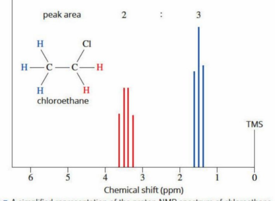
In chloroethane:
- The blue group has 2 neighbouring red hydrogen atoms
- Splitting: (triplet)
- The red group has 3 neighbouring blue hydrogen atoms
- Splitting: (quartet)
The peak area ratio is 3:2, matching the number of hydrogen atoms in each environment.
Worked Example: Methyl propanoate
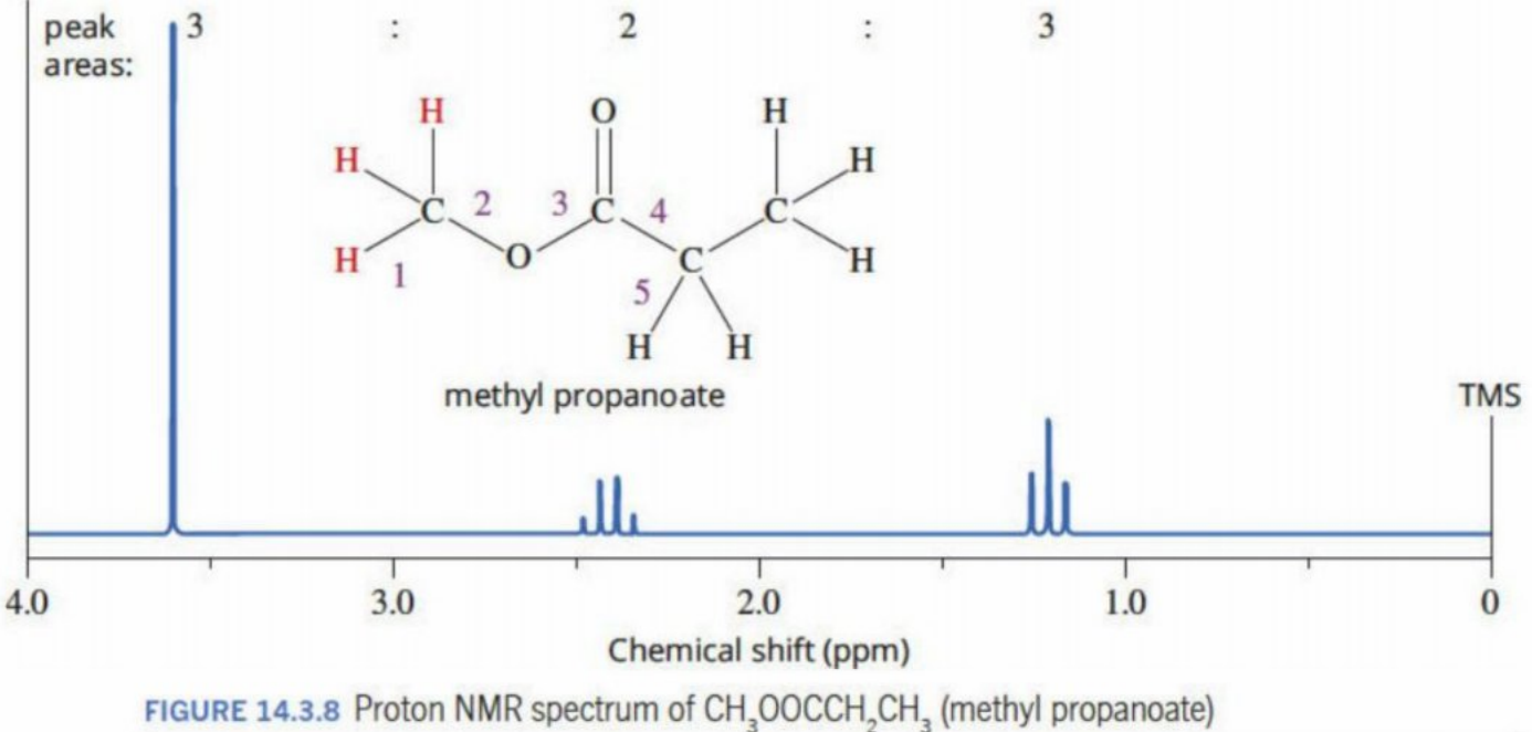
- Red attached to oxygen: more than 3 bonds from other hydrogens → singlet
- Blue : has 3 neighbouring hydrogens in the yellow → quartet
- Yellow : has 2 neighbouring hydrogens in the blue → triplet
Special cases in signal splitting
Equivalent neighbouring atoms don't cause splitting: If neighbouring hydrogen atoms are in the same chemical environment as the hydrogen atoms producing the signal, they won't cause splitting.
Hydroxyl groups: The hydrogen atom in alcohol groups:
- Usually doesn't split neighbouring hydrogen signals
- Usually appears as a singlet itself
- Doesn't count as a neighbour when applying the rule
Multiplets: Sometimes you may see more complex splitting patterns called multiplets. These occur when hydrogen atoms have non-equivalent neighbouring groups, leading to overlapping splitting patterns.
Systematic interpretation of proton NMR spectra
Follow these steps to identify an unknown compound from its proton NMR spectrum:
Step 1: Count the number of signals to determine how many different hydrogen environments exist.
Step 2: Examine the relative peak areas to find the ratio of hydrogen atoms in each environment. Remember to scale this ratio to match the total number of hydrogens in the molecular formula.
Step 3: Analyse the splitting pattern of each signal using the rule to identify neighbouring hydrogen atoms.
Step 4: Use the chemical shift values and Table 14.3.1 to identify the types of functional groups present.
Step 5: Combine all the information to propose a molecular structure. Check that your structure:
- Has the correct molecular formula
- Produces the observed number of signals
- Shows the correct peak area ratios
- Displays the expected splitting patterns
- Has chemical shifts matching the observed values
Worked Example: Identifying Dichloroethane Isomers
Problem: A compound has the molecular formula . Its proton NMR spectrum shows:
- A doublet at 2.1 ppm (relative area = 3)
- A quartet at 5.9 ppm (relative area = 1)
Solution:
Step 1 - Possible structures: Two isomers are possible:
- 1,2-dichloroethane:
- 1,1-dichloroethane:
Step 2 - Analyse the spectrum:
| Chemical shift (ppm) | Peak splitting | Relative peak area |
|---|---|---|
| 2.1 | Doublet | 3 |
| 5.9 | Quartet | 1 |
Step 3 - Number of environments: Two signals = two different hydrogen environments
Step 4 - Number of hydrogens:
- Peak area ratio 1:3 directly gives the number of hydrogens (total = 4)
- One environment has 1 hydrogen, the other has 3 hydrogens
- This indicates a group and a group
Step 5 - Interpret splitting patterns:
- Doublet (2.1 ppm): , so neighbour → this is the group
- Quartet (5.9 ppm): , so neighbours → this is the group
Step 6 - Conclusion: The molecule must be 1,1-dichloroethane () because:
- It has a group adjacent to a group
- The splitting patterns match: the methyl group splits the signal into a quartet, and the splits the methyl signal into a doublet
- 1,2-dichloroethane would show only one signal (all hydrogens equivalent)
Carbon-13 NMR spectroscopy
Principles of carbon-13 NMR
Carbon-13 (C) is a naturally occurring isotope that makes up only 1.1% of all carbon atoms. Unlike carbon-12 (the most abundant isotope), carbon-13 has nuclear spin and can be detected by NMR spectroscopy.
Carbon-13 NMR identifies different carbon atom environments within a molecule. Like proton NMR, it uses TMS as a reference standard (defined as ppm).
Key differences from proton NMR
1. Chemical shift range: Carbon-13 chemical shifts range from 0 to about 220 ppm, much wider than the proton NMR range (0-13 ppm).
2. No splitting patterns: Because carbon-13 is only 1.1% abundant, it's extremely unlikely that two carbon-13 atoms will be next to each other in a molecule. Therefore, carbon-13 NMR spectra don't show splitting by other carbon atoms. All signals appear as single peaks.
3. Peak areas: The peak areas in carbon-13 NMR are NOT directly proportional to the number of carbon atoms in each environment. You cannot use integration to count carbons.
Typical carbon-13 NMR chemical shifts
| Type of carbon | Chemical shift (ppm) |
|---|---|
| 8-25 | |
| 20-45 | |
| 40-60 | |
| 36-45 | |
| (X = F, Cl, Br, I) | 15-80 |
| or | 35-70 |
| 50-90 | |
| (alkyne) | 75-95 |
| (alkene) | 110-150 |
| (carboxylic acid) | 160-185 |
| Ester carbon | 165-175 |
| Ketone/aldehyde carbon | 190-220 |
Study tip: Carbonyl carbons () have the highest chemical shifts, appearing at 160-220 ppm.
Comparing proton and carbon-13 NMR
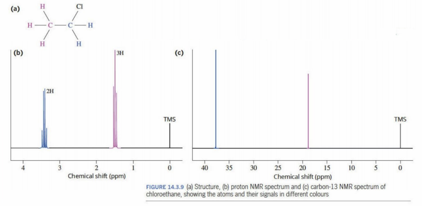
For the same molecule (such as chloroethane shown above):
- Both types of NMR show the same number of signals (equal to the number of different environments)
- Proton NMR shows splitting patterns; carbon-13 NMR doesn't
- Proton NMR peak areas are proportional to hydrogen count; carbon-13 peak areas are not reliable for counting carbons
- Chemical shift ranges are different (0-13 ppm for H, 0-220 ppm for C)
Remember!
Key Points to Remember:
-
NMR spectroscopy uses radio frequency radiation to detect nuclear spin changes in molecules, providing detailed structural information.
-
The number of signals in an NMR spectrum equals the number of different chemical environments. Equivalent atoms (those in the same chemical environment) produce one signal.
-
In proton NMR, four key features help identify structures: (1) number of signals shows hydrogen environments, (2) peak area ratio shows relative number of hydrogens, (3) chemical shift indicates functional groups, and (4) splitting pattern reveals neighbouring hydrogens.
-
The rule states that a signal splits into peaks, where is the number of equivalent neighbouring hydrogen atoms within three bonds. Common patterns: singlet (no neighbours), doublet (1 neighbour), triplet (2 neighbours), quartet (3 neighbours).
-
Carbon-13 NMR shows the number of different carbon environments but differs from proton NMR in three important ways: wider chemical shift range (0-220 ppm), no splitting patterns, and peak areas not proportional to carbon count.