Cell Fractionation & Ultracentrifugation (AQA A-Level Biology): Revision Notes
Cell Fractionation & Ultracentrifugation
What is cell fractionation?
Cell fractionation is a laboratory technique that allows scientists to break apart cells and separate their different organelles into distinct fractions. This process is essential when researchers need to study the structure and function of specific cellular components in isolation, rather than within the complex environment of an intact cell.
To obtain meaningful results from cell fractionation, scientists need large quantities of isolated organelles. This technique has revolutionised our understanding of cellular biology by enabling detailed analysis of what individual organelles actually do.
Cell fractionation is particularly valuable because it allows scientists to study organelles without the interference of other cellular components, providing clearer insights into their specific functions and biochemical processes.
Prerequisites for successful cell fractionation
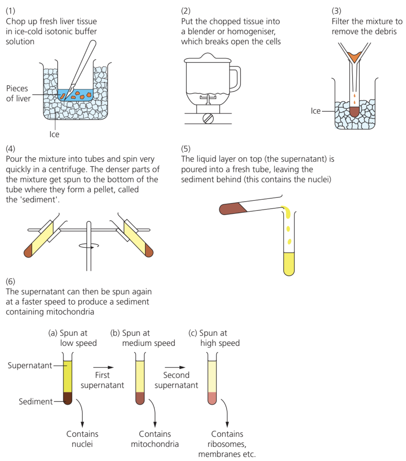
Before beginning cell fractionation, the tissue sample must be placed in a carefully prepared solution with three important characteristics:
Critical Solution Requirements:
The solution must be cold to reduce enzyme activity that could break down and damage the organelles during the separation process. It must have the same water potential as the tissue to prevent organelles from bursting or shrinking due to osmotic water movement. Finally, it must be buffered to maintain a stable pH, since changes in acidity could alter organelle structure or affect enzyme function.
These three conditions - cold temperature, isotonic environment, and pH stability - work together to preserve organelle integrity throughout the fractionation process.
The two main stages
Homogenation
The first stage involves breaking open the cells using a device called a homogeniser or blender. This mechanical disruption releases all the organelles from their cellular compartments into the surrounding solution.
Understanding Homogenation
The resulting mixture, called homogenate, contains all the cellular components floating freely in the buffer solution. However, this mixture also includes any complete cells that weren't broken open and large pieces of cellular debris, which must be removed before proceeding to the separation stage.
The homogenate is filtered to remove these unwanted large particles, leaving behind a solution containing just the isolated organelles ready for separation.
Ultracentrifugation
The second stage uses a machine called a centrifuge to separate the different organelles based on their mass and density. Ultracentrifugation works by spinning tubes of the filtered homogenate at extremely high speeds, creating a powerful centrifugal force.
This force causes the organelles to move through the liquid at different rates - heavier organelles sink faster than lighter ones, allowing them to be collected separately.
Step-by-step separation process
The separation follows a systematic approach, starting with the slowest speed and gradually increasing:
Worked Example: Sequential Organelle Separation
Step 1: The tube containing filtered homogenate is placed in the centrifuge and spun at a relatively slow speed. The heaviest organelles - the nuclei - are forced to the bottom of the tube where they form a solid sediment or pellet.
Step 2: The liquid portion at the top, called the supernatant, is carefully removed and transferred to a fresh tube. This supernatant contains all the remaining organelles except the nuclei.
Step 3: The process continues with the supernatant being spun at a faster speed. This time, the next heaviest organelles - the mitochondria - sediment out at the bottom, while the remaining organelles stay in the new supernatant.
Step 4: This sequential process continues, with each round using higher speeds to separate progressively lighter organelles. The lysosomes require the highest centrifugation speeds to separate effectively.
Centrifugation speeds for different organelles
Different organelles require different centrifugal forces for effective separation:
| Organelle | Centrifugation Speed (revolutions per minute) |
|---|---|
| Nuclei | 1,000 |
| Mitochondria | 3,500 |
| Lysosomes | 16,500 |
These speeds reflect the relative masses of the organelles, with heavier structures requiring less centrifugal force to sediment out of solution. Notice how the speeds increase dramatically as we separate progressively smaller and lighter organelles.
Equipment and practical considerations
The ultracentrifuge is a sophisticated piece of laboratory equipment capable of generating the enormous forces needed to separate cellular components. Modern ultracentrifuges can spin at speeds exceeding 100,000 revolutions per minute, creating forces thousands of times stronger than gravity.
The technique requires careful handling to maintain organelle integrity throughout the process. Temperature control remains important even during centrifugation to prevent enzyme-mediated damage to the separated components.
Significance of cell fractionation techniques
Cell fractionation and ultracentrifugation have enabled major advances in biological knowledge by allowing scientists to study organelles in isolation. This approach revealed detailed information about organelle structure and function that would be impossible to obtain from intact cells.
The technique allows researchers to determine what specific roles different organelles play in cellular processes, leading to our current understanding of cellular metabolism, protein synthesis, and many other biological functions.
Key Points to Remember:
- Cell fractionation separates cellular organelles by first breaking open cells, then using centrifugal force to sort components by mass
- The buffer solution must be cold, buffered, and isotonic to prevent organelle damage during the process
- Homogenation breaks open cells mechanically, while ultracentrifugation separates the released organelles
- Organelles separate in order of decreasing mass: nuclei first, then mitochondria, finally lysosomes
- This technique revolutionised cell biology by enabling detailed study of isolated organelle function