The Electron Microscope (AQA A-Level Biology): Revision Notes
The Electron Microscope
Introduction to electron microscopy
Light microscopes have limited resolution due to the relatively long wavelength of light. In the 1930s, scientists developed a revolutionary alternative that uses a beam of electrons instead of light - the electron microscope. This advancement provided two main advantages over traditional light microscopy.
The electron beam possesses a much shorter wavelength than visible light, enabling the microscope to achieve significantly better resolution. Additionally, since electrons carry a negative charge, they can be precisely focused using electromagnets rather than glass lenses.
Modern electron microscopes can resolve objects separated by just 0.1 nm - approximately 2000 times better resolution than the best light microscopes.
However, electrons interact with air molecules, so specimens must be placed in a near-vacuum environment for the microscope to function effectively.
Types of electron microscope
There are two main categories of electron microscope, each designed for different purposes:
- Transmission electron microscope (TEM)
- Scanning electron microscope (SEM)
Transmission electron microscope
How TEM works
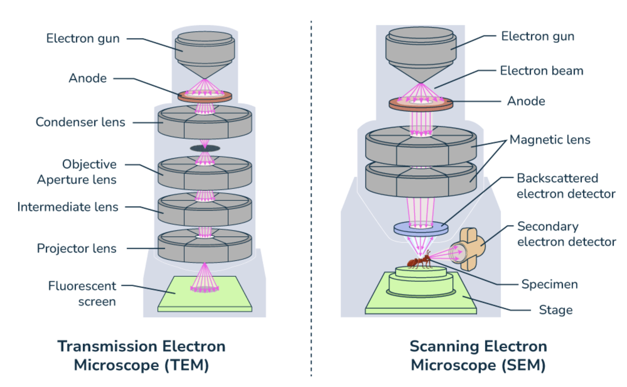
The TEM operates using an electron gun that generates a focused beam of electrons directed onto the specimen via a condenser electromagnet. The electron beam passes through an extremely thin section of the prepared specimen.
Different parts of the specimen interact with electrons differently - some regions absorb electrons and appear dark, while other areas allow electrons to pass through and appear bright. This creates contrast that forms an image on a screen, which can be photographed to produce a photomicrograph.
The contrast in TEM images is created by differential electron absorption - denser areas appear darker while less dense regions appear brighter, similar to how X-rays create contrast in medical imaging.
TEM resolution and limitations
The TEM achieves excellent resolving power of , though this theoretical limit cannot always be reached in practice due to several factors:
Specimen preparation difficulties can limit the achievable resolution, and the higher energy electron beam required may damage delicate specimens.
The main limitations of TEM include:
- Vacuum requirement: The entire system must operate in a vacuum, making it impossible to observe living specimens
- Complex specimen preparation: A lengthy 'staining' process is necessary, and even then the final image lacks colour
- Ultra-thin specimens: Samples must be extremely thin to allow electron penetration
- Artefacts: These are artificial features that may appear during specimen preparation but do not exist in the natural specimen, making it challenging to distinguish genuine structures from preparation artefacts
Artefacts are a critical limitation of electron microscopy. These artificial features can be mistaken for real cellular structures, making it essential for scientists to compare multiple specimens and use different preparation techniques to confirm genuine biological features.
TEM image characteristics
TEM specimens must be cut extremely thin to allow electron penetration, resulting in flat, 2-D images. However, scientists can partially overcome this limitation by taking multiple sections through a specimen and building up a 3-D image from the series of cross-sections.
Scanning electron microscope
How SEM works
The SEM shares many limitations with the TEM, except that specimens do not require extreme thinning since electrons do not need to penetrate the material. Instead, the SEM directs an electron beam onto the specimen surface from above, scanning back and forth across a portion of the specimen in a regular pattern.
The electrons scatter when they hit the specimen surface, and this scattering pattern depends on the surface contours. Computer analysis of the scattered electrons and secondary electrons produced creates detailed images. This technique allows construction of 3-D images showing surface structure.
SEM resolution and capabilities
The basic SEM has lower resolving power than a TEM - around - but this still provides approximately ten times better resolution than light microscopes. The key advantage of SEM is its ability to generate detailed three-dimensional images of surface structures.
Modern computer processing can add false colours to SEM images to enhance contrast and highlight specific features, though the original images are monochrome like TEM images.
Advantages over light microscopy
Electron microscopes offer substantial improvements over light microscopy:
- Superior resolution: Up to 2000 times better than light microscopes
- Enhanced detail: Ability to observe subcellular structures and organelles in unprecedented detail
- Versatile imaging: TEM for internal structures, SEM for surface features
- High magnification: Capable of much higher useful magnification than light microscopes
However, these advantages come with the trade-off of being unable to observe living specimens and requiring complex preparation procedures.
Key Points to Remember:
- Electron microscopes use electron beams instead of light, achieving much better resolution due to shorter wavelengths
- TEM produces 2-D images with resolution by passing electrons through ultra-thin specimens
- SEM creates 3-D surface images with resolution by scanning electrons across specimen surfaces
- Both types require vacuum conditions, preventing observation of living specimens
- Specimen preparation is complex and may introduce artefacts that don't represent natural structures