Homeostasis (AQA A-Level Biology): Revision Notes
Role of the Nephron in Osmoregulation
The kidney maintains the water potential of plasma and tissue fluid through osmoregulation. The nephron performs this vital function through four distinct stages that work together to control water balance and concentrate urine.
The four key stages of nephron function are: ultrafiltration, reabsorption in the proximal convoluted tubule, maintenance of sodium gradients by the loop of Henle, and fine-tuning in the distal convoluted tubule.
Formation of glomerular filtrate by ultrafiltration
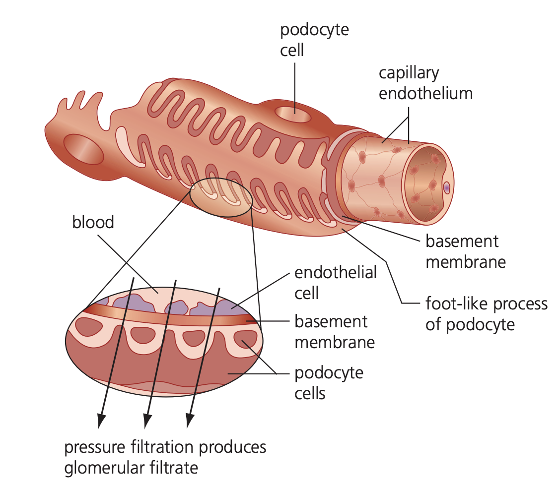
Blood enters the kidney through the renal artery, which branches extensively to form approximately one million tiny arterioles. Each arteriole enters a renal capsule (Bowman's capsule) within a nephron, where it forms a complex network of capillaries called the glomerulus.
The filtration process
The glomerular capillaries merge to form the efferent arteriole, which has a smaller diameter than the afferent arteriole. This size difference creates hydrostatic pressure within the glomerulus, forcing water, glucose, and mineral ions out of the blood to form glomerular filtrate.
Blood cells and proteins cannot pass through the filtration barrier because they are too large. The filtrate formation is assisted by several resistance factors, but two key modifications reduce this resistance:
Specialised Filtration Structures
Podocytes are highly specialised cells forming the inner layer of the renal capsule. These cells have gaps between them, allowing filtrate to pass between rather than through the cells. The endothelium of glomerular capillaries also contains spaces up to 100 nm wide, permitting fluid movement between cells rather than through them.
This design ensures sufficient hydrostatic pressure overcomes resistance, producing approximately 125 cm³ of filtrate per minute containing useful substances that need to be recovered.
Reabsorption of glucose and water by the proximal convoluted tubule
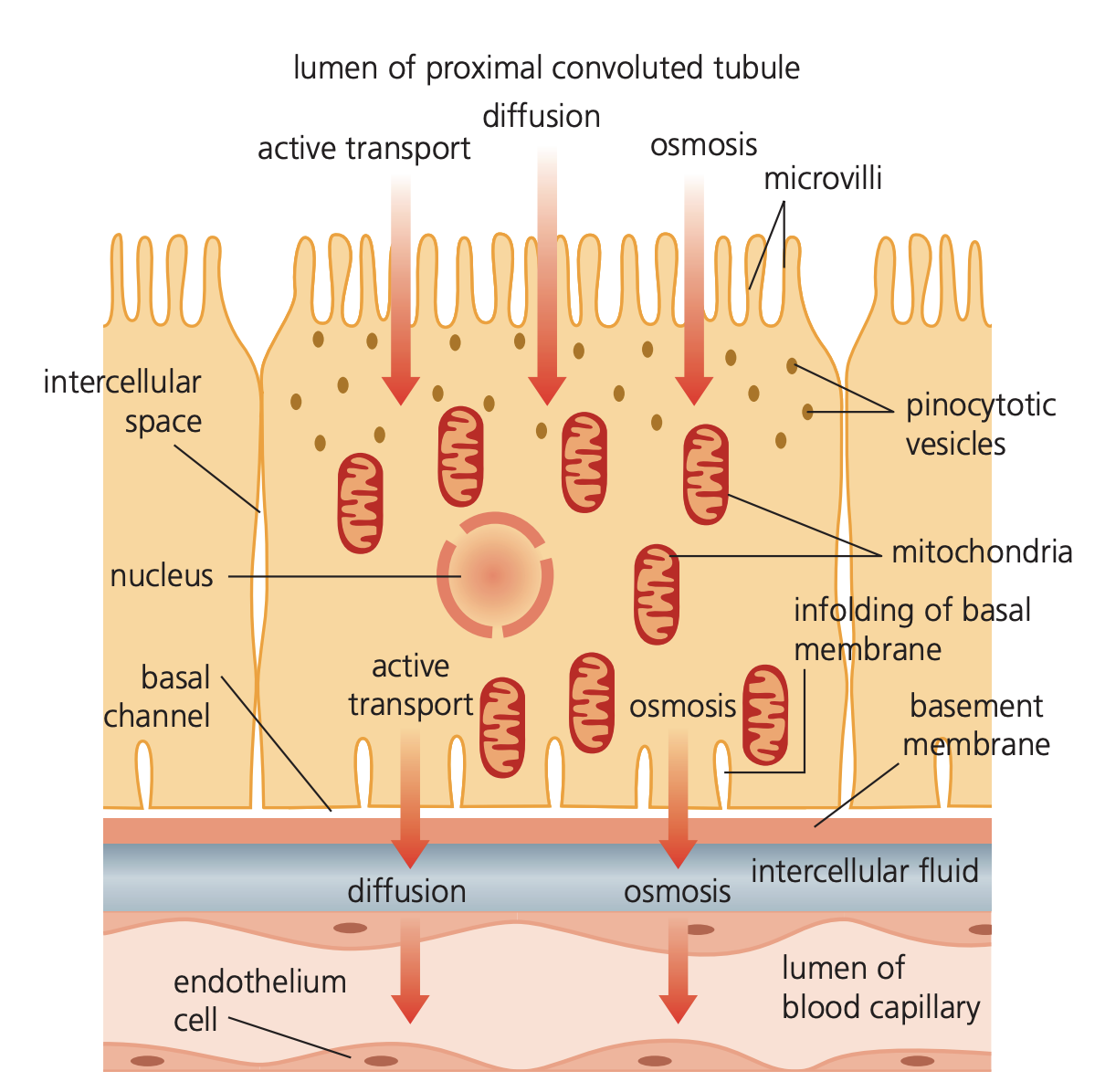
The proximal convoluted tubule (PCT) reabsorbs nearly 85% of the filtrate back into the blood. This process operates through size-based selection - small waste molecules like urea are removed, while useful substances are recovered.
Structural adaptations for reabsorption
PCT cells possess several adaptations that maximise reabsorption efficiency:
- Microvilli provide an extensive surface area for absorbing substances from the filtrate
- Infoldings at the cell bases create large surface areas for transferring reabsorbed materials into blood capillaries
- High mitochondrial density supplies ATP for active transport processes
The reabsorption mechanism
The process follows a specific sequence that demonstrates the elegant efficiency of nephron function:
Worked Example: Co-transport Mechanism in the PCT
Step 1: Sodium ions are actively transported out of PCT cells into blood capillaries, lowering the sodium concentration within the cells
Step 2: Sodium ions then diffuse down their concentration gradient from the tubule lumen into the PCT cells, but only through specialised carrier proteins
Step 3: These carrier proteins use co-transport, simultaneously moving other molecules (glucose, amino acids, chloride ions) along with sodium ions into the cells
Step 4: The co-transported molecules diffuse into the blood, ensuring all glucose and most other valuable substances are reabsorbed along with water
This system recovers essential nutrients while allowing waste products to continue through the nephron for elimination.
Maintenance of a gradient of sodium ions by the loop of Henle
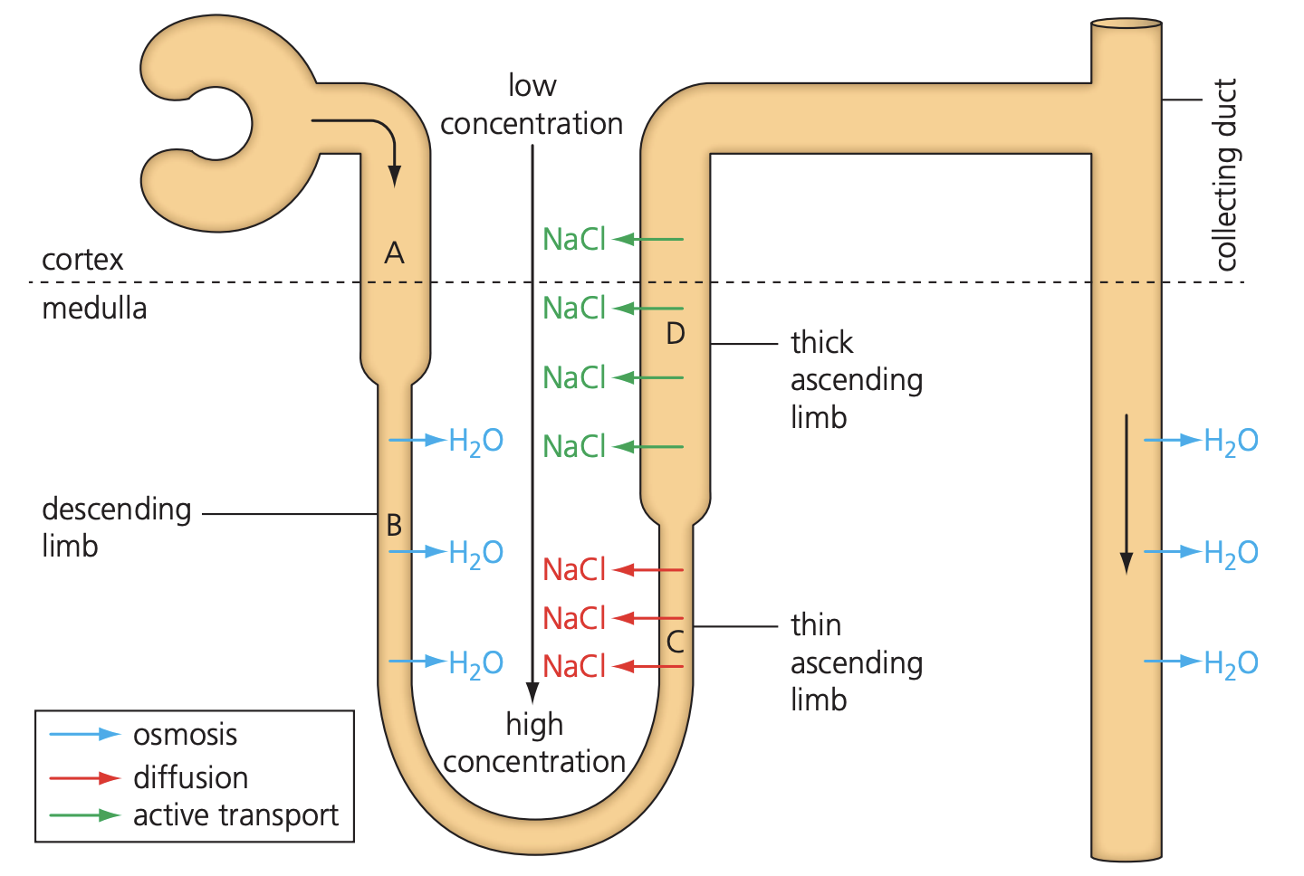
The loop of Henle is a hairpin-shaped structure extending into the kidney's medulla. It creates a concentration gradient that enables water reabsorption from the collecting duct, producing concentrated urine with lower water potential than blood.
Structure and function
The loop contains two distinct regions with contrasting properties:
- Descending limb: narrow with thin walls that are highly permeable to water
- Ascending limb: wider with thick walls that are impermeable to water
The contrasting permeabilities of the descending and ascending limbs are essential for creating the concentration gradient. If both limbs had the same permeability, the counter-current multiplier system would not function effectively.
The counter-current multiplier mechanism
This system works as a counter-current multiplier because the filtrate flows in opposite directions in the two limbs, enhancing the concentration effect:
Worked Example: Counter-Current Multiplier Process
Step 1: Active transport removes sodium ions from the ascending limb using ATP from numerous mitochondria in the wall cells
Step 2: This creates low water potential (high ion concentration) in the medulla between the two limbs - the interstitial region
Step 3: Water passes out of the descending limb by osmosis into this concentrated interstitial space, then enters blood capillaries
Step 4: As filtrate moves down the descending limb, it progressively loses water, lowering its water potential
Step 5: Sodium ions diffuse out at the base of the ascending limb, and are actively pumped out as the filtrate moves up, creating progressively higher water potential
Step 6: A water potential gradient develops in the interstitial space, with the highest water potential in the cortex and lowest in the medulla
Step 7: Water passes from the collecting duct by osmosis as the filtrate moves through this gradient
Step 8: The counter-current flow maintains a constant water potential gradient, ensuring continuous water extraction from the filtrate
About 180 dm³ of water enters the nephrons daily, but only 1 dm³ leaves as urine, demonstrating the remarkable efficiency of this reabsorption system.
The distal convoluted tubule
The distal convoluted tubule performs fine adjustments to water and salt balance. Its cells contain microvilli and numerous mitochondria, enabling rapid material reabsorption through active transport.
Fine-Tuning Function
The main function involves making precise adjustments to water and salt levels in response to the body's needs. The tubule walls can alter their permeability under hormonal control, allowing selective reabsorption of ions while controlling pH through selective ion reabsorption.
This final stage ensures the filtrate composition is precisely adjusted before reaching the collecting duct, where antidiuretic hormone (ADH) can further regulate water loss through aquaporins - specialised water channel proteins.
Key Points to Remember:
- Ultrafiltration creates filtrate through hydrostatic pressure, with podocytes and endothelial gaps facilitating the process
- The proximal convoluted tubule reabsorbs 85% of filtrate using active transport and co-transport mechanisms powered by ATP
- The loop of Henle operates as a counter-current multiplier, creating a concentration gradient that enables water reabsorption from the collecting duct
- Structural adaptations throughout the nephron (microvilli, mitochondria, specialised permeabilities) maximise the efficiency of each process
- The system processes 180 dm³ of water daily but produces only 1 dm³ of urine, demonstrating remarkable conservation efficiency