Structure of the Nephron (AQA A-Level Biology): Revision Notes
Structure of the Nephron
Overview of kidney structure
The kidney contains approximately one million microscopic tubular structures called nephrons, which serve as the basic structural and functional units. Each kidney is organised into distinct regions: the outer cortex and inner medulla, both containing different parts of the nephron structure.
The kidney's organisation into cortex and medulla regions allows for the specialised functions of different nephron components to occur in optimal environments.
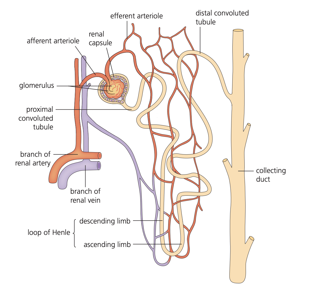
The kidney connects to the body's circulation through the renal artery (delivering blood from the heart) and renal vein (returning blood to circulation). Urine produced by nephrons collects in the renal pelvis before passing through the ureter to the bladder.
Basic nephron structure
Each nephron forms a narrow tube approximately 14mm in length, sealed at one end and featuring two convoluted (twisted) regions connected by a hairpin-shaped loop. The nephron consists of five main structural components arranged in sequence.
Understanding that the nephron is a sealed tube is crucial - this closed system allows for the precise control of filtration and reabsorption processes.
Components of the nephron
Renal (Bowman's) capsule
The renal capsule, also known as Bowman's capsule, forms the closed starting point of each nephron. This cup-shaped structure surrounds a dense cluster of blood capillaries called the glomerulus.
The inner wall of the renal capsule contains specialised cells called podocytes, which have unique structural features that allow them to form filtration barriers around the glomerular capillaries.
Podocytes are highly specialised cells with foot-like projections that wrap around glomerular capillaries, creating precisely sized filtration slits.
Proximal convoluted tubule
Following the renal capsule, the proximal convoluted tubule forms a series of twisted loops surrounded by numerous blood capillaries. The walls of this section consist of epithelial cells equipped with microvilli - tiny projections that significantly increase the surface area for absorption and secretion processes.
Loop of Henle
The Loop of Henle creates a distinctive hairpin-shaped structure that extends from the cortex down into the medulla and back up again. This loop comprises two sections:
- Descending limb - carries fluid down into the medulla
- Ascending limb - returns fluid back towards the cortex
The entire loop remains surrounded by blood capillaries throughout its length.
The hairpin shape of the Loop of Henle is essential for creating concentration gradients that allow the kidney to produce concentrated urine.
Distal convoluted tubule
The distal convoluted tubule forms another series of twisted loops, similar to the proximal section but with important structural differences. Its walls consist of epithelial cells, but these contain fewer capillaries surrounding them compared to the proximal tubule.
Collecting duct
Multiple distal convoluted tubules from several nephrons connect to a single collecting duct. This tube gradually widens as it progresses towards the renal pelvis, where it empties the processed filtrate. The collecting duct is lined with epithelial cells and becomes increasingly wide as it approaches the kidney's central cavity.
Blood supply to the nephron
Afferent arteriole
The afferent arteriole represents a tiny blood vessel that branches from the renal artery system. This vessel delivers blood directly to the nephron by entering the renal capsule, where it forms the glomerular capillary network.
Glomerulus
The glomerulus consists of a tightly packed, many-branched cluster of capillaries contained within the renal capsule. This arrangement creates a unique capillary bed where an arteriole supplies blood and another arteriole drains it - unusual compared to typical capillary beds drained by venules.
The glomerulus is unique in the circulatory system because it's a capillary bed between two arterioles rather than between an arteriole and a venule. This arrangement is crucial for maintaining filtration pressure.
Efferent arteriole
The efferent arteriole forms a tiny vessel that carries blood away from the renal capsule after it has passed through the glomerular capillaries. This vessel has a smaller diameter than the afferent arteriole, creating important pressure differences within the glomerulus.
Capillary networks
Beyond the glomerulus, the efferent arteriole branches extensively to form blood capillaries that create dense networks surrounding the proximal convoluted tubule, Loop of Henle, and distal convoluted tubule. These capillaries eventually merge into venules and then into the renal vein system.
Structural organisation
The nephron demonstrates remarkable structural organisation with each component positioned to facilitate specific processes. The renal capsule and glomerulus occupy the cortex region, while the Loop of Henle extends into the medulla. The proximal and distal convoluted tubules remain within the cortex, surrounded by extensive capillary networks.
The strategic positioning of nephron components across cortex and medulla regions allows for the creation of concentration gradients essential for water conservation.
This structural arrangement ensures that each nephron maintains close contact with blood supply throughout its length, while the collecting ducts from multiple nephrons converge to channel processed filtrate towards the ureter system.
Key Points to Remember:
- Each kidney contains approximately one million nephrons - the functional units consisting of a sealed tube with two twisted regions
- The renal (Bowman's) capsule contains specialised podocytes and surrounds the glomerulus capillary cluster
- The proximal convoluted tubule features microvilli to increase surface area, while the Loop of Henle creates a hairpin structure extending into the medulla
- Blood flows from afferent arteriole → glomerulus → efferent arteriole → surrounding capillary networks
- The collecting duct receives filtrate from multiple nephrons and gradually widens as it approaches the renal pelvis