Structure of Synapses (AQA A-Level Biology): Revision Notes
Structure of Synapses
What are synapses?
A synapse represents the communication junction between one neurone and another neurone, or between a neurone and an effector organ. These structures play a vital role in linking different neurones and coordinating nervous system activities throughout the body.
Unlike electrical impulses that travel along neurones, synapses transmit information through chemical messengers, allowing for more precise and controlled neural communication.
Unlike electrical impulses that travel along neurones, synapses transmit information through chemical messengers called neurotransmitters. This chemical transmission system allows precise control over neural communication.
Components of synaptic structure
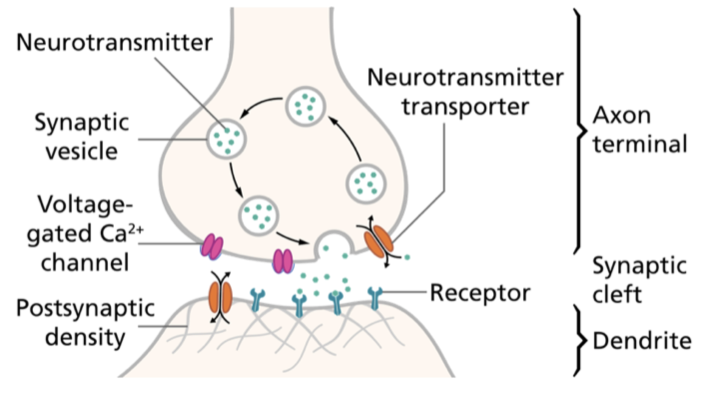
The synaptic gap
Neurones do not physically connect at synapses. Instead, they are separated by a narrow space called the synaptic cleft, which measures approximately 20-30 nanometres in width.
This tiny gap of 20-30 nanometres is incredibly small - about 200 times smaller than the width of a human hair! This precise spacing ensures that information transfer occurs through controlled chemical release rather than direct electrical contact.
Presynaptic neurone
The neurone that sends the signal is termed the presynaptic neurone. Its axon terminates in a swollen region called the synaptic knob, which contains several important structures:
- Mitochondria - provide energy for neurotransmitter synthesis and release
- Smooth endoplasmic reticulum - manufactures neurotransmitters
- Synaptic vesicles - store neurotransmitters ready for release
- Calcium ion protein channels - control neurotransmitter release when opened
Postsynaptic neurone
The receiving neurone is called the postsynaptic neurone. Its membrane contains:
- Receptor proteins - bind specifically to neurotransmitters
- Sodium ion protein channels - open when neurotransmitters bind to receptors
- These channels consist of five protein subunits that form the complete channel structure
Key features of synaptic function
Unidirectionality
Synapses operate as one-way valves for information flow. Signals can only pass from the presynaptic neurone to the postsynaptic neurone, never in reverse.
This directional control is essential for organised neural pathways. Without it, signals could travel backwards and create chaos in the nervous system.
This occurs because:
- Only presynaptic terminals contain neurotransmitter-filled vesicles
- Only postsynaptic membranes possess the appropriate receptor proteins
- This directional control ensures organised neural pathways
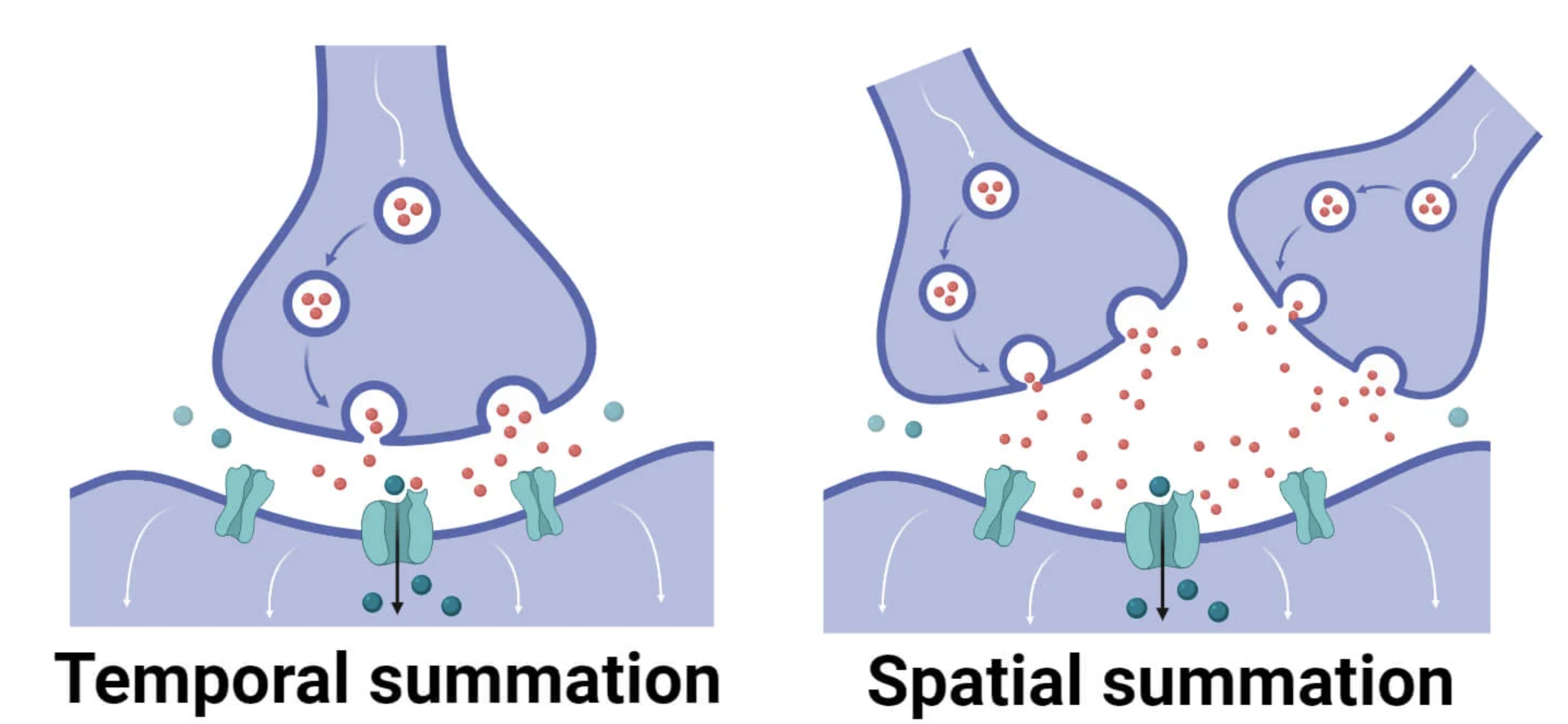
Summation processes
Individual synaptic transmissions often release insufficient neurotransmitter to trigger new action potentials in postsynaptic neurones. Summation solves this problem by building up neurotransmitter concentrations through two distinct mechanisms:
Worked Example: Spatial Summation
Imagine three different presynaptic neurones (A, B, and C) all connecting to the same postsynaptic neurone. Each releases a small amount of neurotransmitter:
- Neurone A releases 30% of the threshold amount
- Neurone B releases 35% of the threshold amount
- Neurone C releases 40% of the threshold amount
When all three fire simultaneously: 30% + 35% + 40% = 105% of threshold This exceeds the 100% needed, so an action potential is generated in the postsynaptic neurone.
Spatial summation occurs when multiple different presynaptic neurones simultaneously release neurotransmitter at the same postsynaptic neurone. The combined chemical release from several sources creates sufficient concentration to exceed the postsynaptic threshold value, generating a new action potential.
Worked Example: Temporal Summation
A single presynaptic neurone fires repeatedly in quick succession:
- First release: 40% of threshold (insufficient alone)
- Second release (before first breaks down): 40% + 35% = 75% (still insufficient)
- Third release: 75% + 30% = 105% (exceeds threshold)
The rapid succession prevents complete neurotransmitter breakdown between releases, allowing accumulation to reach threshold.
Temporal summation involves a single presynaptic neurone releasing neurotransmitter repeatedly over a short time period. Rapid, successive releases allow neurotransmitter concentration to accumulate before breakdown occurs, again reaching threshold levels in the postsynaptic neurone.
Both summation types ensure reliable signal transmission even when individual synaptic events produce sub-threshold responses.

Inhibitory synapses
Not all synapses promote action potential generation. Inhibitory synapses reduce the likelihood of postsynaptic action potentials through a different neurotransmitter mechanism:
- Presynaptic neurones release inhibitory neurotransmitters
- These bind to chloride ion protein channels on postsynaptic membranes
- Chloride ions (Cl⁻) enter the postsynaptic neurone through facilitated diffusion
- This binding also opens nearby potassium ion (K⁺) protein channels
- Potassium ions move out of the postsynaptic neurone into the synapse
Understanding Hyperpolarisation
The combined effect creates hyperpolarisation - think of it as making the neurone "extra negative" and therefore much harder to excite. It's like raising the bar higher for action potential generation.
The combined effect creates hyperpolarisation - the membrane potential becomes more negative (approximately compared to the usual resting potential). This increased negativity makes action potential generation much more difficult, as larger sodium ion influxes are needed to reach threshold.
Key Points to Remember:
- Synapses use chemical transmission - neurotransmitters cross the synaptic cleft to carry information between neurones
- Information flows in one direction only - from presynaptic to postsynaptic neurone, ensuring organised neural pathways
- Summation mechanisms amplify weak signals - spatial summation uses multiple neurones whilst temporal summation uses rapid repeated releases
- Inhibitory synapses prevent unwanted signals - they use chloride and potassium ion movements to hyperpolarise postsynaptic membranes
- Synaptic structure supports function - vesicles store neurotransmitters while specific receptor proteins ensure selective binding