Control of Heart Rate (AQA A-Level Biology): Revision Notes
Control of Heart Rate
The autonomic nervous system
The autonomic nervous system manages self-governing processes within the body. This system controls involuntary activities of internal muscles and glands without conscious input. The autonomic system coordinates responses to internal stimuli, ensuring our body systems adapt efficiently to changing demands.
The autonomic nervous system operates completely outside of conscious control, managing vital functions like heart rate, breathing, and digestion automatically.
The autonomic nervous system operates through two distinct divisions that work in opposition:
Sympathetic nervous system - This division generally stimulates effectors and accelerates activity. It functions like an emergency controller, preparing the body for stressful situations by heightening awareness and readiness for action (the fight or flight response). When activated, it increases physiological processes to cope with demanding circumstances.
Parasympathetic nervous system - This division typically inhibits effectors and reduces activity levels. It manages activities during normal resting conditions, focusing on conserving energy and replenishing the body's reserves. This system dominates when the body is at rest.
These two systems are antagonistic, meaning their actions normally oppose each other. When one system contracts a muscle, the other causes relaxation. This balanced opposition allows precise regulation of internal organs and muscles.
Understanding the antagonistic relationship between sympathetic and parasympathetic systems is crucial - they work in opposition to provide fine control over heart rate and other vital functions.
Basic heart rate control mechanism
The heart muscle, known as cardiac muscle, differs from other muscles because it is myogenic. This means contractions originate from within the muscle tissue itself, rather than requiring nervous impulses from outside sources.
Located within the right atrium wall is a specialised group of cells called the sinoatrial node (SAN). This structure generates the initial stimulus for heart contractions and establishes the heart's basic rhythm. The SAN functions as the heart's natural pacemaker, determining the fundamental heart rate through its regular pattern of electrical stimulation.
The myogenic nature of cardiac muscle means the heart can continue beating even when removed from the body, as long as it has oxygen and nutrients. This is why hearts can be transplanted successfully.
The electrical conduction pathway follows a specific sequence:
- A wave of electrical excitation spreads from the sinoatrial node across both atria, causing them to contract
- A layer of non-conductive tissue (atrioventricular septum) prevents the wave from crossing directly to the ventricles
- The electrical wave enters the atrioventricular node (AVN), positioned between the atria
- After a brief delay, the AVN conveys the electrical wave to the ventricles through specialised muscle fibres called Purkyne tissue, which collectively form the bundle of His
- The bundle of His conducts the wave through the atrioventricular septum to the base of the ventricles
- The wave spreads into smaller Purkyne fibres throughout the ventricle walls
- Electrical excitation releases from the Purkyne tissue, causing both ventricles to contract simultaneously from the bottom upwards
Modifying resting heart rate
A typical adult human maintains a resting heart rate of approximately 70 beats per minute. However, this rate must adapt to meet varying oxygen demands, such as during exercise when the heart rate may need to more than double.
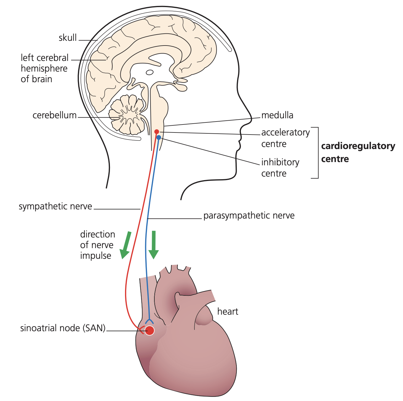
Heart rate modifications are controlled by a brain region called the medulla oblongata. This structure contains two distinct centres responsible for heart rate regulation:
- A centre that increases heart rate, connected to the sinoatrial node via the sympathetic nervous system
- A centre that decreases heart rate, linked to the sinoatrial node through the parasympathetic nervous system
The medulla oblongata acts as the control centre for heart rate regulation, processing input from various receptors and coordinating appropriate responses through the autonomic nervous system.
The activation of these centres depends on nerve impulses received from two types of receptors that monitor blood chemistry and pressure changes.
Control by chemoreceptors
Chemoreceptors are located within the walls of the carotid arteries (arteries supplying the brain). These receptors detect pH changes in blood that result from alterations in carbon dioxide concentration. When carbon dioxide dissolves in blood, it forms an acid, thereby lowering the blood's pH.
The chemoreceptor control mechanism operates as follows:
During increased muscular or metabolic activity, tissues produce more carbon dioxide through enhanced respiration. This increased carbon dioxide production lowers blood pH below normal levels.
Chemoreceptors in the carotid arteries detect this pH reduction and increase the frequency of nerve impulses sent to the medulla oblongata. Specifically, they stimulate the centre responsible for increasing heart rate.
This centre responds by increasing impulse frequency via the sympathetic nervous system to the sinoatrial node. The SAN consequently increases its rate of electrical wave production, elevating the heart rate.
The elevated heart rate increases blood flow, which removes carbon dioxide more rapidly through the lungs. As carbon dioxide concentration returns to normal, blood pH rises back to normal levels.
When pH normalises, chemoreceptors in the carotid arteries reduce their impulse frequency to the medulla oblongata. The medulla oblongata then decreases impulses to the sinoatrial node, leading to heart rate reduction.
This process creates a negative feedback loop that maintains blood pH within normal ranges during varying activity levels. The system automatically corrects deviations from the normal pH range.
Control by pressure receptors
Pressure receptors are positioned within the walls of both the carotid arteries and the aorta. These receptors monitor blood pressure changes and trigger appropriate heart rate adjustments.
When blood pressure exceeds normal levels, pressure receptors increase nerve impulses to the medulla oblongata centre that decreases heart rate. This centre sends impulses via the parasympathetic nervous system to the sinoatrial node, reducing the heart's beating rate and thereby lowering blood pressure.
When blood pressure falls below normal levels, pressure receptors increase nerve impulses to the medulla oblongata centre that increases heart rate. This centre sends impulses via the sympathetic nervous system to the sinoatrial node, accelerating the heart rate and increasing blood pressure.
This pressure receptor system provides another negative feedback mechanism, ensuring blood pressure remains within appropriate ranges for efficient circulation.
Integration of control systems
The autonomic nervous system integrates multiple inputs from both chemoreceptors and pressure receptors to maintain cardiovascular homeostasis. The medulla oblongata processes information from these various sources and coordinates appropriate responses through both sympathetic and parasympathetic pathways.
This integrated control allows the cardiovascular system to respond effectively to changing physiological demands while maintaining stable internal conditions. The antagonistic relationship between sympathetic and parasympathetic divisions ensures precise regulation of heart rate under varying circumstances.
The integration of multiple control systems allows for precise cardiovascular regulation. The body can simultaneously respond to changes in blood chemistry, pressure, and metabolic demands to maintain optimal circulation.
Key Points to Remember:
-
The autonomic nervous system controls heart rate through sympathetic (increases rate) and parasympathetic (decreases rate) divisions that work antagonistically
-
The sinoatrial node (SAN) acts as the heart's natural pacemaker, with electrical signals conducted through AVN, bundle of His, and Purkyne fibres
-
The medulla oblongata contains separate centres for increasing and decreasing heart rate based on receptor input
-
Chemoreceptors detect blood pH changes from CO₂ levels and adjust heart rate to maintain acid-base balance during exercise
-
Pressure receptors monitor blood pressure and trigger heart rate changes to maintain appropriate circulation pressure through negative feedback mechanisms