Photoreceptors (AQA A-Level Biology): Revision Notes
Photoreceptors
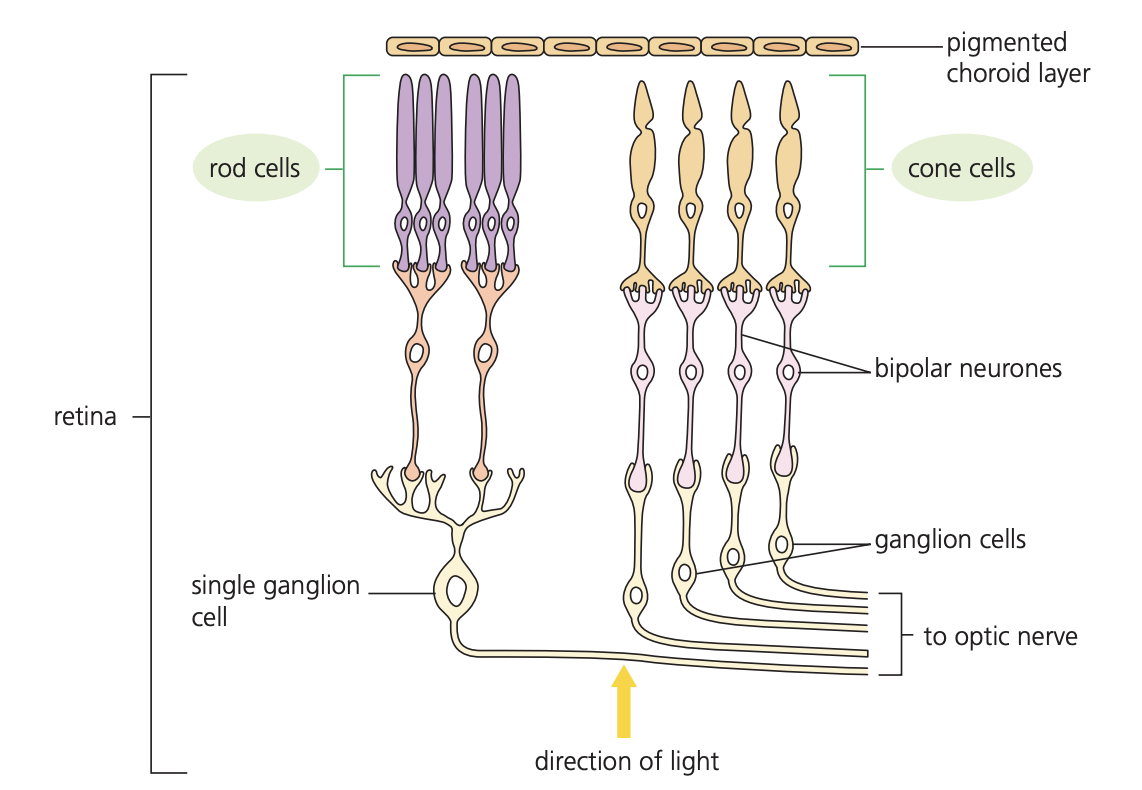
Photoreceptors are specialised sensory cells found in the retina of the mammalian eye. These cells function as transducers, converting light energy into electrical energy that can be transmitted as nerve impulses to the brain. There are two main types of photoreceptors: rod cells and cone cells, each with distinct characteristics and functions.
Rod cells
Rod cells are one of the two main types of photoreceptors in the mammalian retina. These cells are specifically adapted for detecting light in low-intensity conditions, making them essential for vision in dim environments.
Structure and numbers
Rod cells have an elongated, rod-like shape and are extremely numerous in the human eye. Each eye contains approximately 120 million rod cells, making them far more abundant than cone cells. The high number of rod cells reflects their important role in low-light vision.
Function and sensitivity
Rod cells are highly sensitive to light and can detect very low light intensities. This sensitivity allows humans to see in dim conditions, such as at night. However, this comes with a trade-off - rod cells cannot distinguish between different wavelengths of light. As a result, vision provided by rod cells appears only in black and white, without colour discrimination.
The high sensitivity of rod cells is achieved through a process called retinal convergence. Multiple rod cells connect to a single bipolar cell, which then connects to a sensory neurone. This arrangement means that even if only a few rod cells are stimulated by low-intensity light, their combined signals can exceed the threshold needed to generate a generator potential in the connected bipolar cell.
Visual acuity
While rod cells excel at detecting low-intensity light, they provide relatively poor visual acuity. This means that fine detail cannot be distinguished clearly when relying primarily on rod cell vision. The convergence of many rod cells to single bipolar cells means that the brain cannot pinpoint exactly which rod cells were stimulated, resulting in less precise spatial information.
Cone cells
Cone cells are the second type of photoreceptor, specialised for high-intensity light detection and colour vision. These cells are cone-shaped and far less numerous than rod cells.
Structure and numbers
Each human eye contains approximately 6 million cone cells. While this is significantly fewer than rod cells, cone cells provide crucial capabilities for daytime vision and colour perception.
Types and colour vision
Cone cells exist in three distinct types, each containing different photosensitive pigments called iodopsin. Each type responds optimally to different wavelengths of light, enabling colour vision. The three types detect different ranges of the light spectrum, and the brain interprets the relative stimulation of these different cone types to produce the perception of colour.
Function and sensitivity
Unlike rod cells, cone cells require high light intensities to function effectively. The pigment iodopsin in cone cells needs more energy to break down compared to rhodopsin in rod cells. This means cone cells only respond when sufficient high-intensity light is available, explaining why colour vision is poor in dim lighting conditions.
Each cone cell typically connects to its own individual bipolar cell, rather than sharing connections like rod cells. This one-to-one relationship allows for much greater precision in detecting the exact location of light stimuli.
Visual acuity
The individual connections between cone cells and bipolar cells, combined with their concentrated distribution, give cone cells excellent visual acuity. This allows for the perception of fine detail and sharp images when adequate light is available.
Distribution on the retina
The distribution of rod and cone cells across the retina is uneven and reflects their different functions.
The fovea
The fovea is a small area of the retina where light is focused by the lens. This region has the highest concentration of cone cells but contains no rod cells. The high density of cone cells at the fovea, combined with their individual connections to bipolar cells, provides the sharpest vision and best colour discrimination.
Peripheral regions
Rod cells are distributed mainly around the periphery of the retina, with very few present at the fovea. This distribution explains why objects at the edge of our visual field are detected more easily in dim light, but appear in black and white and lack fine detail.
The concentration of cone cells decreases significantly moving away from the fovea towards the periphery of the retina, while rod cell density increases.
Working together
The combination of rod and cone cells provides mammals with excellent vision across a wide range of lighting conditions. During bright daylight, cone cells dominate vision, providing sharp, coloured images. As light levels decrease, rod cells become increasingly important, allowing continued vision albeit with reduced colour perception and visual acuity.
This dual system explains many aspects of human vision, such as why peripheral vision is more sensitive to movement in low light, and why looking slightly to the side of a dim star makes it easier to see than looking directly at it.
Key Points to Remember:
-
Rod cells are numerous (120 million per eye), detect low-intensity light, provide black and white vision, and have poor visual acuity due to convergence onto bipolar cells
-
Cone cells are fewer (6 million per eye), require high-intensity light, exist in three types for colour vision, and provide excellent visual acuity through individual bipolar cell connections
-
Rod cells concentrate at the retinal periphery while cone cells concentrate at the fovea, creating different visual capabilities across the visual field
-
Both cell types act as transducers, converting light energy into electrical nerve impulses that travel to the brain
-
The distribution and properties of these photoreceptors explain why we see colours and fine detail best in bright light, but can detect movement and objects better at the edge of our vision in dim conditions