Mitosis (OCR A-Level Biology A): Revision Notes
Mitosis
Introduction to mitosis
Mitosis is the type of nuclear division that produces two genetically identical daughter cells. This process occurs when new cells are needed for:
- Growth of tissues and organisms
- Repair of damaged tissues
- Asexual reproduction in eukaryotic organisms
The mother cell (original cell) must first duplicate its chromosomes so that each daughter cell receives a complete set of genetic information.
Although mitosis is described using distinct stages for convenience, the actual process flows continuously with each stage blending smoothly into the next. There are no pauses between stages—the division is one seamless progression.
Interphase and the cell cycle
Interphase is not a stage of mitosis itself, but rather the period of the cell cycle when the cell is not dividing. During interphase, several preparations occur:
- DNA replication takes place during the S (synthesis) phase
- The DNA exists as uncoiled chromatin, making it invisible under a light microscope
- Organelles duplicate in preparation for cell division
- Centrioles replicate in animal cells
By the time mitosis begins, each chromosome already consists of two identical DNA molecules. This duplication is essential for producing genetically identical daughter cells.
Stages of mitosis
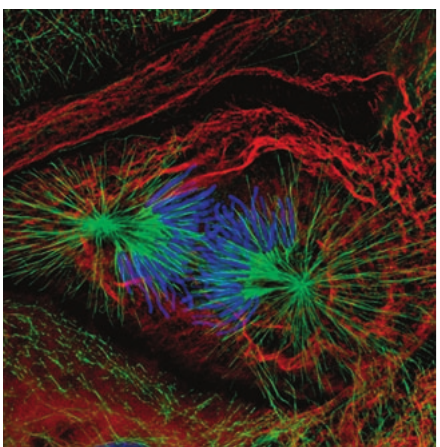
The process of mitosis is divided into four main stages: prophase, metaphase, anaphase, and telophase.
Prophase
Prophase marks the beginning of visible chromosome formation. During this stage:
- Chromosomes become visible because DNA undergoes supercoiling (the twisting of DNA around its own axis, winding the helix more tightly)
- Each chromosome appears as two sister chromatids joined at a single point called the centromere
- The nucleolus disappears
- The nuclear membrane breaks down by the end of prophase
At this point, each chromosome contains two DNA molecules as a result of earlier replication during interphase. The chromatids remain joined at the centromere throughout prophase and metaphase.
Metaphase
During metaphase, the cell prepares to separate the sister chromatids:
- In animal cells, centrosomes (each containing a pair of duplicated centrioles) move to opposite ends of the cell, called poles
- The centrosomes extend microtubules that form the spindle apparatus, creating a framework for chromatid movement
- Chromosomes attach to spindle fibres at the cell's centre (the equator)
- Attachment occurs via protein structures called kinetochores located on each chromatid at the centromere
Anaphase
Anaphase is the stage where sister chromatids separate:
- Centromeres divide, releasing the sister chromatids
- The newly-separated chromatids (now called chromosomes) are pulled toward opposite poles
- Spindle fibres shorten at both ends, pulling the chromatids apart
Critical terminology: When a chromosome consists of two sister chromatids joined at the centromere, each part is called a chromatid. Once they separate during anaphase, each individual chromatid becomes a chromosome. By telophase, you should refer to these structures as chromosomes rather than chromatids.
Telophase
Telophase represents the final stage of nuclear division:
- New nuclear membranes form around each group of chromosomes at opposite poles
- Chromosomes begin to uncoil, returning to their extended chromatin state
- New nucleoli form within each nucleus
At the end of telophase, the cell contains two nuclei, each with a complete set of chromosomes identical to the original cell.
Cytokinesis
After telophase, the cytoplasm divides in a process called cytokinesis. Although not technically part of mitosis (which refers only to nuclear division), cytokinesis completes the formation of two separate cells.
In animal cells:
- The cytoplasm near the equator pinches inward
- The plasma membrane tucks in, eventually splitting the cytoplasm
- Organelles, which increased in number during interphase, are distributed between the two cells
- The Golgi apparatus produces vesicles that fuse to form new sections of plasma membrane
Mitosis in plants
Mitosis in plant cells follows the same basic stages as in animal cells, with two key differences:
Key Differences Between Plant and Animal Mitosis:
-
Absence of centrioles: Plant cells lack centrioles but still form a functional spindle apparatus from microtubules
-
Different cytokinesis: Instead of the membrane pinching inward, vesicles from the Golgi apparatus migrate to the equator and fuse to form:
- The new cell membrane (plasma membrane)
- The new cell wall
Mitosis and asexual reproduction
Asexual reproduction is reproduction involving the production of offspring from a single individual, without the fusion of gametes. In eukaryotic organisms, asexual reproduction always involves mitosis to generate the new cells.
Key characteristics of asexual reproduction:
- Produces organisms that are genetically identical to the parent
- All offspring are clones of each other and the parent
- The only genetic variation arises from spontaneous mutations
Advantages and Disadvantages of Asexual Reproduction:
Advantages:
- Much faster than sexual reproduction
- No need to find a mate (particularly useful for sessile organisms like plants)
- Successful genotypes are preserved
Disadvantages:
- Lack of genetic variation reduces adaptability
- If one organism is susceptible to disease or environmental change, all individuals will be equally vulnerable
- Entire populations may be eliminated by a single threat
Asexual reproduction is far more common in plants than animals, as plants cannot move to find mates.
Budding in yeast
Yeast, a unicellular fungus, reproduces asexually through a process called budding. This process uses mitosis for nuclear division but differs from typical cell division:
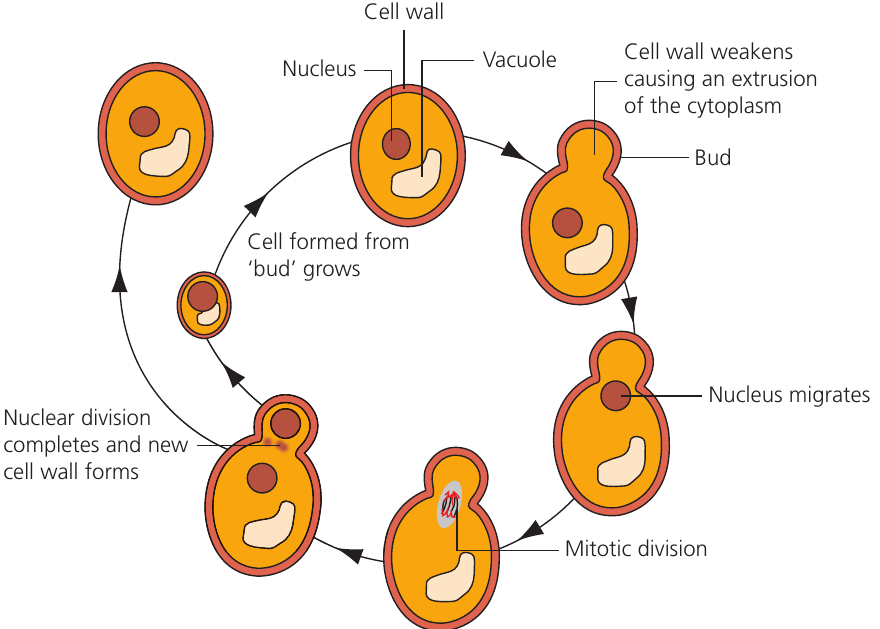
Stages of budding:
- DNA replicates inside the nucleus during interphase
- An area of the cell wall weakens; turgor pressure causes the cytoplasm and cell wall to bulge outward, forming a bud
- A ring of chitin forms at the junction between the parent cell and bud (this remains as a 'scar' after separation)
- The nucleus migrates toward the bud region and undergoes mitotic division (the nuclear membrane remains intact throughout, unlike in animal and plant cells)
- One daughter nucleus migrates into the bud along with organelles
- Cytokinesis completes with formation of a new cell wall between mother and daughter cells
- The daughter cell grows to full size before beginning its own budding cycle
Key Differences from Typical Mitosis:
- Unequal cytoplasmic division: The mother cell retains most of its cytoplasm, while the bud receives less
- Nuclear membrane: Remains intact during nuclear division
- Growth pattern: Only the daughter cell (bud) needs to grow; the mother cell is already full-sized and can begin budding again immediately
In contrast, animal and plant cell division produces two daughter cells of equal size, both smaller than the original, and both must grow to reach full size.
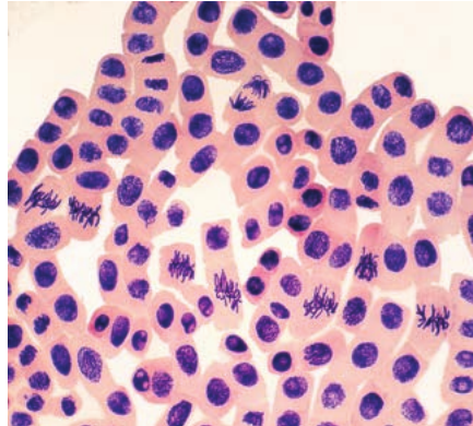
Observing mitosis and the mitotic index
Mitosis can be observed in actively dividing tissues such as root tips. The mitotic index provides a quantitative measure of cell division activity:
Practical observation method
Root tips from garlic or onion can be prepared for microscopic observation:
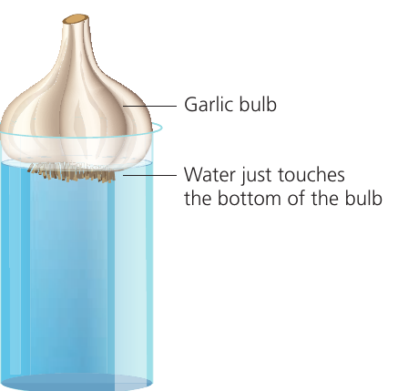
Key preparation steps:
- Grow roots from a garlic bulb with its base just touching water
- Cut cm root tips
- Fix cells using ethanoic acid to stop all cellular processes
- Treat with M hydrochloric acid at to break down cell wall bonds (allowing cells to separate)
- Stain chromosomes using ethano-orcein stain
- Squash the tissue to create a thin, single-cell layer
- Observe under a microscope and count cells at each stage
Sample data analysis
| Stage | Number of cells seen |
|---|---|
| Prophase | |
| Metaphase | |
| Anaphase | |
| Telophase | |
| Interphase | |
| Total |
Worked Example: Calculating Mitotic Index
From the data above, we can calculate the mitotic index:
Step 1: Count cells undergoing mitosis Cells in mitosis = Prophase + Metaphase + Anaphase + Telophase Cells in mitosis = cells
Step 2: Apply the formula
Step 3: Interpret the stages
- Prophase represents the longest stage (most cells observed: )
- Anaphase is the shortest stage (fewest cells observed: )
The duration of each stage affects the number of cells observed at that stage. Longer stages will show more cells, as each cell spends more time in that phase.
Temporal variation in mitotic activity
Cell division rates can vary throughout the day due to biological rhythms:
| Time | Mitotic index (%) |
|---|---|
| 12 midnight | |
| 4 am | |
| 8 am | |
| 12 noon | |
| 4 pm | |
| 8 pm |
This data shows peak mitotic activity at midday () and lowest activity at midnight (), suggesting circadian regulation of cell division.
Cell cycle timing example
Different cell types have different cycle durations. For bone marrow cells with an -hour cycle:
- Interphase: of the cycle ( hours hours)
- Mitosis: of the cycle ( hours hours)
- Cytokinesis: of the cycle ( hours hours)
Remember!
Key Points to Remember:
-
Mitosis produces two genetically identical daughter cells with the same chromosome number as the parent cell, essential for growth, repair, and asexual reproduction
-
The four stages (PMAT) are prophase (chromosomes condense), metaphase (chromosomes align at equator), anaphase (chromatids separate), and telophase (nuclei reform)
-
Interphase is not part of mitosis but is when DNA replicates during S phase, preparing the cell for division
-
Plant and animal mitosis differ slightly: plants lack centrioles and use Golgi vesicles to form cell walls during cytokinesis
-
Mitotic index measures division activity and is calculated as (cells in mitosis ÷ total cells) × 100%, useful for comparing division rates in different tissues or conditions