Tissues, Organs, and Organ Systems (OCR A-Level Biology A): Revision Notes
Tissues, Organs, and Organ Systems
Introduction to tissues
In multicellular organisms, cells are organised into tissues. A tissue is a group of cells working together to carry out a particular function. While cells in a tissue are often similar, they may include more than one cell type.
Blood is an excellent example of a tissue containing multiple cell types—it includes distinct red blood cells and several types of white blood cells, all working together to carry out blood's various functions.
Animal tissues
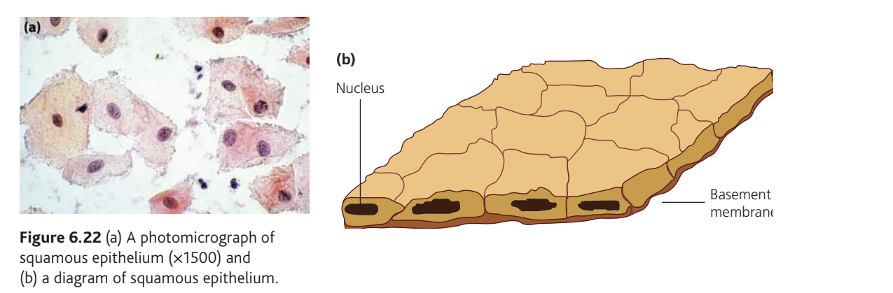
Squamous epithelium
Squamous epithelium refers to any epithelial tissue consisting of flattened cells arranged in a layer. This tissue is found throughout the animal body and sits on a basement membrane.
Epithelium is a sheet of cells forming an outer layer—a covering or lining of body surfaces.
Basement membrane is a thin, delicate membrane composed of protein fibres and polysaccharides that separates epithelium from underlying tissue.
Structure and function
Simple squamous epithelium consists of a single layer of relatively unspecialised but flattened cells. Because these cells form a surface covering, their thin profile reduces the distance substances must travel to pass through the tissue, thereby shortening the diffusion pathway.
The flattened shape of squamous epithelial cells is a critical adaptation. By being thin, these cells create the shortest possible diffusion pathway, enabling efficient exchange of gases, nutrients, and waste products across the epithelial barrier.
Ciliated epithelium
Ciliated epithelium is composed of cells bearing numerous cilia on their surfaces. This tissue is found in animals where material needs to be moved across a surface—for instance, in the trachea (to move mucus) and in the oviduct (to move the ovum).
Structure and function
The coordinated beating of cilia shifts material along the epithelial surface. These cells are highly specialised for this movement function. Key structural features include:
- Cilia: hair-like projections from the cell surface
- Basal granules: spherical bodies at the base of cilia and flagella
- Cytoplasm: containing cellular machinery
- Nucleus: controlling cell activities
- Basement membrane: supporting the epithelial layer
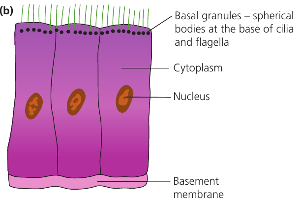
The cilia in the trachea beat in a coordinated wave-like pattern, moving mucus and trapped particles upward and away from the lungs. This is an essential protective mechanism for the respiratory system.
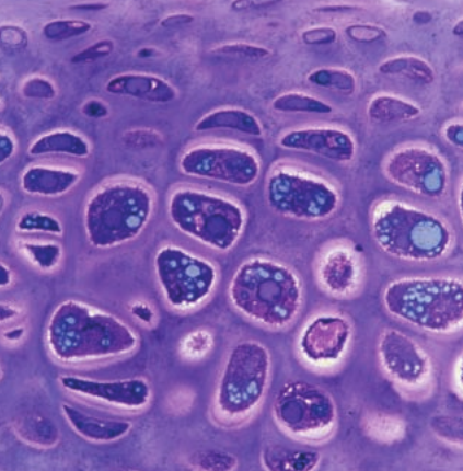
Cartilage
Cartilage is an example of connective tissue—tissue that connects, supports, binds, or separates other tissues or organs.
Structure and composition
Cartilage is made up of specialised cells called chondrocytes, which produce an extracellular matrix containing:
- Collagen fibres: provide stiffness and strength
- Elastin fibres: provide flexibility
The balance between collagen and elastin fibres determines the properties of different types of cartilage. Areas requiring more flexibility (like the ear) have more elastin, while areas needing strength (like joints) have more collagen.
Function and location
The primary function of cartilage is to protect and strengthen. In mammals, cartilage is found in:
- Nose
- Ear
- Rib cage
- Trachea and bronchi
- Joints
Muscle
Muscle is a highly unusual tissue because its function is to move parts of the body. A muscle (such as the biceps) is actually an organ composed mostly of muscle tissue along with nerve tissue, blood vessels, and connective tissue.
Types of muscle
Mammals possess three types of muscle:
- Skeletal muscle (voluntary)
- Smooth muscle (involuntary)
- Cardiac muscle (found only in the heart)
Hierarchical organisation
The organisation of muscle tissue is complex and hierarchical:
- Myofilaments: contractile proteins called actin and myosin
- Myofibrils: structures formed from myofilaments
- Muscle fibres (cells): highly specialised, multinucleate cells containing myofibrils
- Fascicles: groups of muscle fibres
- Muscle: groups of fascicles, plus nerve fibres, blood vessels, and connective tissue
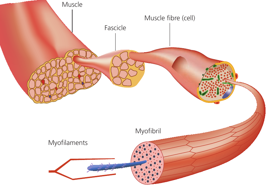
Skeletal muscle cells are commonly referred to as muscle fibres due to their elongated shape. A critical feature is that they are multinucleate, meaning they contain multiple nuclei within each cell. This adaptation supports the high metabolic demands of muscle contraction.
Plant tissues
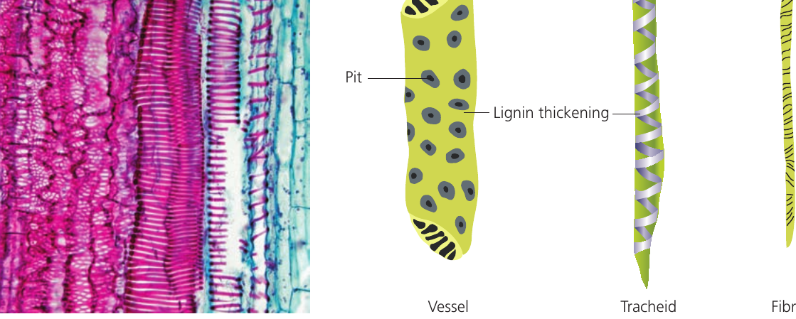
Xylem
Xylem is a plant tissue that transports water and minerals upward through the plant stem and also provides structural support. It may contain up to four different cell types (the exact composition varies between species):
- Vessel elements
- Tracheids
- Fibres
- Parenchyma
Cell types and their functions
Parenchyma cells are relatively unspecialised and form packing tissue between the other cells.
Vessel elements are the main water-transporting cells. They possess:
- A wide lumen (internal space)
- Perforated or completely absent end walls
- Pits (thin areas) allowing lateral water movement, helping to bypass blockages
- Alignment forming continuous tubes running the length of the plant
Tracheids also transport water. In angiosperms (a large group of plants that produce flowers and seeds), they additionally provide structural support. Compared to vessels, tracheids have:
- A narrower lumen
- Perforated end walls and pits for water movement between cells
Fibres do not transport water; their sole function is support. They consist of:
- Minimal or no lumen
- Essentially strips of lignin
Lignification
The walls of vessel elements, tracheids, and fibres are all thickened with lignin, strengthening the walls and enabling their support function. Lignin thickening may appear as:
- Rings
- Spirals
- A more or less continuous sheet perforated by pores
The pattern of lignin deposition varies depending on the location and age of the xylem tissue. Ring and spiral patterns allow the xylem to stretch as the plant grows, while continuous sheets provide maximum strength in mature tissues.
Dead cells
All xylem cells (vessels, tracheids, and fibres) are dead at maturity and lack cytoplasm. This absence of cytoplasm is unnecessary for structural support and would obstruct water transport.
The fact that xylem cells are dead at maturity is a crucial adaptation. The absence of cytoplasm creates an unobstructed pathway for water flow and eliminates the metabolic needs of living cells, making water transport more efficient.
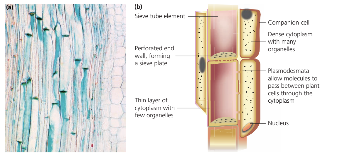
Phloem
Phloem is the plant tissue responsible for transporting organic nutrients (such as sugars) both upward and downward through the plant. Like xylem, it consists of multiple cell types:
- Sieve tube elements
- Companion cells
- Phloem parenchyma (packed between the other cells)
Sieve tube elements
Sieve tube elements are the cells that transport nutrients. To facilitate this function, they have:
- Reduced cytoplasm
- Few organelles
- End walls forming sieve plates—perforated structures allowing cytoplasm of adjacent cells to connect
Companion cells
The reduction in cytoplasm means sieve tube elements cannot maintain themselves independently and require support from companion cells. While the exact role of companion cells remains under investigation, their large numbers of mitochondria and ribosomes suggest they play an active part in phloem transport.
Plasmodesmata are cytoplasmic connections that allow molecules to pass between adjacent plant cells through their cytoplasm. These connections are especially important in phloem tissue, linking sieve tube elements with their companion cells.
Organs and organ systems
Organs
An organ is a group of different tissues that work together. In both animals and plants, specialised organs perform particular functions. These functions often have different aspects requiring different tissues.
Animal Organ Example: The Heart
The heart pumps blood around the body and requires multiple tissue types:
- Cardiac muscle: for contraction and pumping
- Blood vessels: to supply nutrients and oxygen for respiration
- Nervous tissue: to coordinate the beating of the heart
Each tissue contributes a specific function, and all must work together for the heart to function successfully.
Plant Organ Example: The Leaf
The leaf carries out photosynthesis and gas exchange and contains:
- Xylem: brings water to the leaf
- Phloem: transports organic nutrients away from the leaf
- Palisade and spongy parenchyma: carry out photosynthesis
- Epidermis: waterproofs the leaf and contains stomata for gas diffusion
The integration of these different tissues allows the leaf to perform its complex functions efficiently.
Organ systems
In animals, organs often work together to form organ systems. An example is the digestive system, which involves multiple organs:
- Oesophagus
- Stomach
- Small and large intestines
- Liver
- Pancreas
- Gall bladder
Each organ in the digestive system has a specialised role, but they must all work in coordination to break down food, absorb nutrients, and eliminate waste. This demonstrates how organ systems represent a higher level of biological organisation.
Remember!
Key Points to Remember:
-
Tissues are groups of cells working together for a specific function and may contain more than one cell type.
-
Epithelial tissues (squamous and ciliated) are adapted to their functions—squamous epithelium has thin, flattened cells reducing diffusion distance, while ciliated epithelium uses cilia to move materials.
-
Plant transport tissues are highly specialised—xylem cells are dead with lignified walls for water transport and support, while phloem sieve tube elements have reduced cytoplasm and work with companion cells to transport organic nutrients.
-
Muscle tissue has a hierarchical structure from myofilaments (actin and myosin) up to the whole muscle organ.
-
Organs are combinations of different tissues working together, and in animals, multiple organs can form organ systems such as the digestive system.