Microscopy (Edexcel GCSE Biology Combined Science): Revision Notes
Microscopy
Cells are studied using microscopes
Magnifiers use lenses to:
- Magnify images
- Increase the resolution of the image (clearer & more detail)
Advances in Microscope Technology and Understanding of Cell Structures
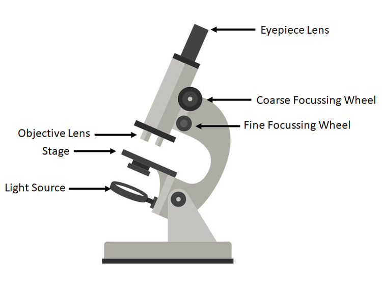
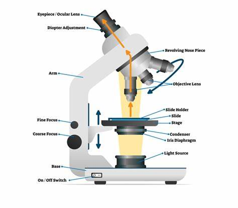
Light Microscopes
Invention and Use:
Light microscopes were invented in the 16th century and were the first tools used to observe cells. They use light to magnify objects, allowing us to see cell structures that are invisible to the naked eye.
Magnification and Resolution:
Light microscopes typically magnify up to about 1500 times (1500x), but their resolution (ability to see detail) is limited to about 200 nanometers (nm). This means we can see larger structures like the nucleus and cell membrane, but smaller organelles, such as ribosomes and detailed internal structures of mitochondria, remain blurry.
Limitation:
The limited resolution of light microscopes meant that smaller sub-cellular structures couldn't be seen clearly, leading to an incomplete understanding of cell function.
Electron Microscopes
Introduction of Electron Microscopy:
In the 1930s, the development of electron microscopes revolutionised cell biology. Instead of using light, these microscopes use a beam of electrons to create an image. This drastically improves resolution and magnification.
Transmission Electron Microscope (TEM):
TEM allows scientists to view the internal structure of cells at an extremely high resolution (up to 0.2 nm), which is over a thousand times better than a light microscope. This enabled us to see organelles like the endoplasmic reticulum, Golgi apparatus, and the internal structure of mitochondria with great detail.
Scanning Electron Microscope (SEM):
SEM gives detailed 3D images of the surface of cells. It helps in visualising the external structure of cells and how organelles are arranged.
Impact on Cell Understanding:
Electron microscopes enabled scientists to discover and understand organelles that were previously unseen.
For example:
- The detailed structure of the mitochondria, showing the inner folds called cristae, helped explain how these organelles generate energy.
- The discovery of ribosomes helped us understand their role in protein synthesis, essential for all cellular processes.
- Chloroplasts in plants were understood in greater detail, especially how their internal membranes carry out photosynthesis.
Advances in Knowledge and Medical Research
Clarity and Detail:
Electron microscopy provided the first clear images of organelles like lysosomes, vesicles, and ribosomes. Understanding these organelles in greater detail has led to breakthroughs in understanding how cells function and communicate.
Medical Research:
Improved microscope technology has advanced medical research by helping scientists understand how viruses and bacteria invade cells, how cancer cells differ from healthy cells, and how cells communicate and respond to different treatments.
3D Imaging:
Modern microscopes, like the confocal laser scanning microscope, allow for 3D reconstructions of cells, helping scientists see how structures are organised and interact within cells.
| Electron microscopes | Light microscopes |
|---|---|
| Invented in 1950s Work by passing light through the specimen Can see nuclei & chloroplasts & study living cells | Invented in 1930s Higher magnification & resolution so can see smaller things in more detail like internal structures Mitochondria & chloroplasts & how cell works & roll of subcellular structures |