Structure of a Synovial Joint (OCR GCSE Physical Education): Revision Notes
📚 Revision Notes
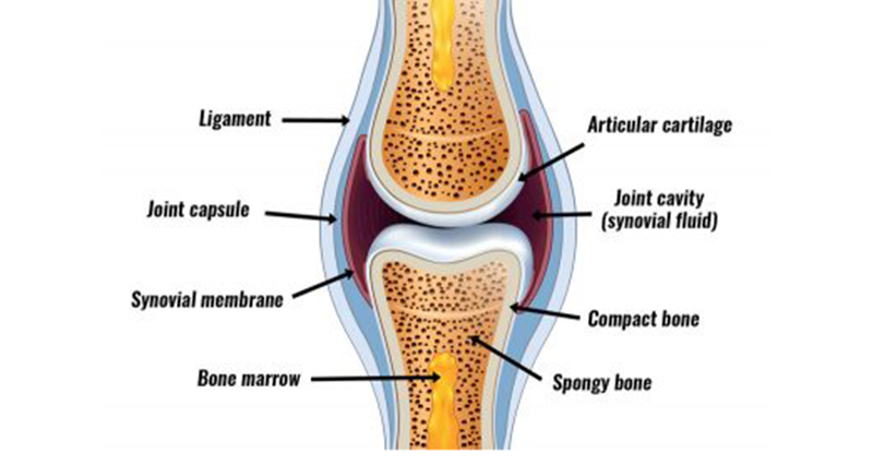
Structure Of A Synovial Joint
infoNote
Bursae: Fluid filled sacs that provides a cushion between the tendons and bones reducing friction
Key Components of a Synovial Joint
- Articular Cartilage: A smooth, slippery tissue that covers the ends of bones within the joint. It reduces friction and absorbs shock during movement.
- Synovial Membrane: A thin lining on the inner surface of the joint capsule. It secretes synovial fluid, which lubricates the joint to facilitate smooth movement.
- Synovial Fluid: A thick, slippery fluid produced by the synovial membrane. It reduces friction, nourishes the cartilage, and helps absorb shocks.
- Joint Capsule: A tough, fibrous structure that encloses the joint cavity. It provides stability and contains the synovial membrane and fluid.
- Ligaments: Strong, flexible bands of connective tissue that connect bones to other bones. They provide stability to the joint by preventing excessive movements.
- Tendons: Fibrous tissues that attach muscles to bones. They help in transmitting the force generated by muscles to move the bones at the joint.
- Bursa: A small fluid-filled sac located between moving structures (e.g., where tendons pass over bones). It reduces friction and allows smooth movement.
- Menisci (or Articular Discs): C-shaped pieces of cartilage in some synovial joints (e.g., knee). They act as shock absorbers and improve the fit between bone ends.