Protein Structure and Function (VCE SSCE Biology): Revision Notes
Protein Structure and Function
Introduction to proteins
Proteins are large, complex biomacromolecules that are essential to all living organisms. A protein is a biomacromolecule made of amino acid chains folded into a three-dimensional shape. Proteins are also known as polypeptides, which are long chains of amino acids. Some proteins consist of a single polypeptide chain, whilst others are made up of multiple polypeptide chains bonded together.
Proteins are one of the four major types of biomacromolecules found in living things. They play crucial roles in virtually every process that occurs within cells and organisms, from providing structural support to catalysing chemical reactions.
Proteins are essential to all life processes. Without properly functioning proteins, cells cannot maintain their structure, respond to their environment, catalyse reactions, or carry out any of the complex processes necessary for survival.
The functional diversity of proteins
Proteins demonstrate remarkable functional diversity, meaning they can perform many different roles within organisms. Understanding this diversity is important because proteins rarely work alone. Instead, they interact with each other to form complex structures and carry out intricate biological processes.
The proteome refers to all the proteins that are expressed by a cell or organism at a given time. Studying the proteome is particularly important to researchers because it provides insight into how proteins work together in living systems.
Eight main functions of proteins
Proteins serve at least eight major functions in living organisms:
1. Enzymes
Enzymes are organic molecules, typically proteins, that catalyse (speed up) specific chemical reactions in the body. An enzyme is an organic molecule that catalyses specific reactions without being consumed in the process. Examples include:
- Catalase, which breaks down hydrogen peroxide into water and oxygen
- Amylase, a digestive enzyme that breaks down starch into maltose
- RNA polymerase, which catalyses the formation of mRNA from DNA
2. Transport proteins
These proteins are typically embedded in cell membranes and control the entry and exit of substances from cells. Examples include:
- Chloride channels
- Glucose channels
- Sodium-potassium pumps
3. Structural proteins
Structural proteins provide support and shape to cells and tissues. Examples include:
- Keratin, a tough protein found in skin, hair, and nails
- Elastin, found in elastic connective tissues such as skin
- Collagen, found in connective tissues such as tendons and ligaments
4. Defence proteins
These proteins are involved in the immune system, recognising and destroying pathogens. An antibody (also known as immunoglobulin) is a protein produced by plasma cells during the adaptive immune response that is specific to an antigen and combats pathogens in various ways. Examples include:
- Antibodies
- Complement proteins
5. Motor and contractile proteins
These proteins are involved in muscle contraction and movement, the movement of internal cell contents around the cytoplasm, and the movement of cilia and flagella. Examples include:
- Myosin and actin, which work together to enable muscle contraction
- Kinesin, which moves along microtubules, enabling mitosis and vesicular transport
6. Storage proteins
Storage proteins act as reserves for metal ions and other molecules throughout organisms. Examples include:
- Ferritin, which stores iron
- Casein, which stores amino acids, carbohydrates, and minerals
7. Receptor proteins
Receptor proteins receive signals from the environment. Examples include:
- Acetylcholine receptors
- Hormone receptors
8. Hormones
Many peptide hormones are protein signalling molecules that regulate physiology or behaviour. These chemical messengers are used to communicate and induce changes in cells. Examples include:
- Insulin, which regulates blood sugar levels
- Adrenaline, which increases heart rate and expands airways
Remembering protein functions:
A helpful mnemonic is "Every Tiger Seems Dangerous, Maybe Some Really Hungry" - representing Enzymes, Transport, Structural, Defence, Motor/contractile, Storage, Receptors, and Hormones. Whilst you don't need to memorise specific protein examples, understanding the range of functions is essential.
Amino acid structure
Amino acids are the fundamental building blocks of proteins. Understanding their structure is key to understanding how proteins are formed and how they function.
Basic components of amino acids
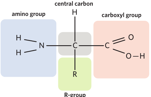
Each amino acid molecule has a consistent basic structure built around a central carbon atom. This central carbon is bonded to four different components:
- A hydrogen atom (H)
- A carboxyl group (COOH) - the functional group that contains a hydroxyl group (OH) and an oxygen double-bonded to a carbon atom
- An amino group (NH₂) - the functional group made up of one nitrogen and two hydrogens
- An R-group (also called a side chain) - the variable portion that differs between amino acids
Memory aid for amino acid components:
Remember "CHAR + C" - Central carbon, Hydrogen, Amino group, R-group, and Carboxyl group. These five components are present in every amino acid.
The R-group and amino acid diversity
The R-group is the variable portion of an amino acid molecule. It can be one of twenty variations and determines the identity of the amino acid. This means there are 20 different types of amino acids in existence, each with its own unique R-group.
The R-group not only identifies the amino acid but also determines its chemical properties. This explains why proteins are not only made up of carbon (C), hydrogen (H), oxygen (O), and nitrogen (N) atoms, but can also contain other elements such as sulphur (S), depending on which R-groups are present.
The R-group is crucial to protein function. It determines:
- The identity of each amino acid
- The chemical properties of the amino acid
- How different amino acids will interact with each other
- How the protein will ultimately fold into its functional shape
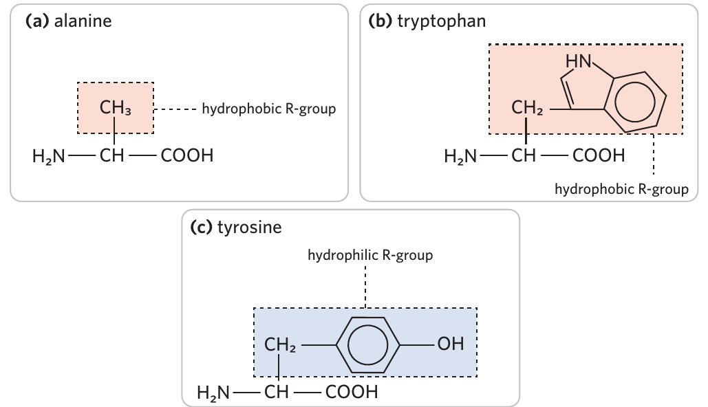
Hydrophobic and hydrophilic properties
Each R-group has specific chemical properties that affect how different amino acids interact with each other within a protein. One important distinction is whether an R-group is hydrophobic or hydrophilic:
- Hydrophobic means having a tendency to repel and be insoluble in water
- Hydrophilic means having a tendency to be attracted to and dissolve in water
An amino acid with a hydrophobic R-group is more likely to form bonds with other amino acids containing hydrophobic R-groups than with amino acids containing hydrophilic R-groups. These interactions are crucial in determining how proteins fold into their functional shapes.
Formation of polypeptides
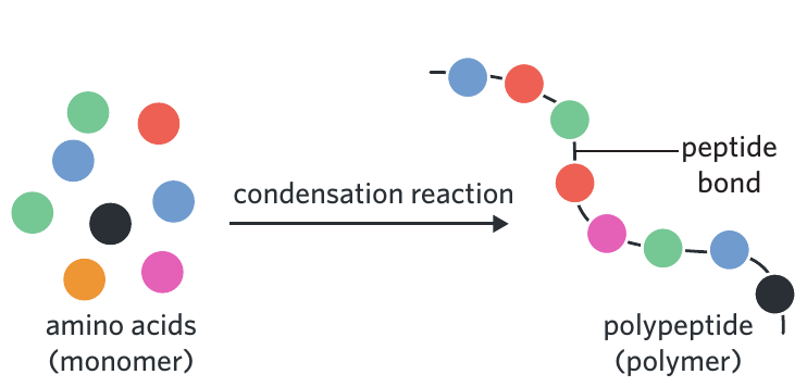
When amino acids join together, they form long chains called polypeptide chains or proteins. Because amino acids have similar basic structures and can act as repeating subunits, they are classified as monomers. A monomer is a molecule that is the smallest building block of a polymer.
When monomers join together, they form polymers. A polymer is a large molecule made up of small, repeated monomer subunits. Therefore, polypeptides and proteins are polymers of amino acids.
Condensation reactions and peptide bonds
The joining of amino acids occurs at a cell's ribosomes through a condensation reaction, which is a reaction where two monomers join to form a larger molecule, producing water as a by-product.
When amino acids join through a condensation reaction, a peptide bond forms between them. A peptide bond is the chemical bond linking two amino acids. These peptide bonds connect adjacent amino acids along the polypeptide chain.
Exam tip: You should be able to draw a generalised diagram of an amino acid showing the central carbon, hydrogen atom, carboxyl group, amino group, and R-group. You don't need to memorise the 20 different amino acids and their specific R-groups.
Protein structure
For a protein to function correctly, the polypeptide chain (or chains) must fold into the correct shape. Protein structure is described in four hierarchical levels, each building upon the previous one. These levels range from the simple sequence of amino acids to complex three-dimensional structures.
Memory aid for structure levels:
Remember "Please Stop Talking Quietly" - Primary, Secondary, Tertiary, Quaternary. This represents the four hierarchical levels of protein structure.
Primary structure
The primary structure is the first level of protein structure, which refers to the sequence of amino acids in a polypeptide chain. This is simply the specific order in which amino acids are arranged along the chain.
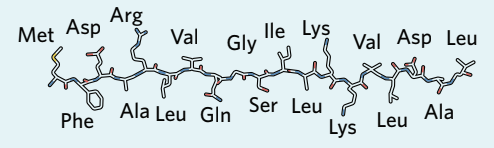
The primary structure is fundamental because it determines all higher levels of protein structure. The sequence of amino acids dictates how the protein will fold and, ultimately, what function it will perform.
Secondary structure
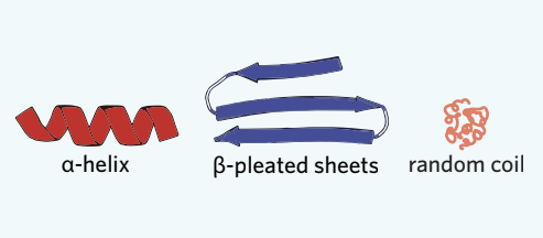
The secondary structure is the level of protein structure where the amino acid chain forms either alpha-helices, beta-pleated sheets, or random coils.
Secondary structure forms when the polypeptide chain folds and coils through the formation of hydrogen bonds between amino acids in different sections of the chain. This folding creates characteristic patterns:
- Alpha helix (α-helix): An organised coiled secondary structure of proteins that resembles a spiral staircase
- Beta-pleated sheet (β-sheet): An organised folded secondary structure of proteins that forms a sheet-like arrangement
- Random coil: An irregular secondary structure of proteins that is neither an alpha helix nor a beta-pleated sheet. Random coils typically join alpha-helices and beta-pleated sheets together
Memory aid for secondary structures:
Think "ABR" - Alpha helices, Beta-pleated sheets, Random coils. These three types of secondary structures are connected by hydrogen bonds between different parts of the polypeptide chain.
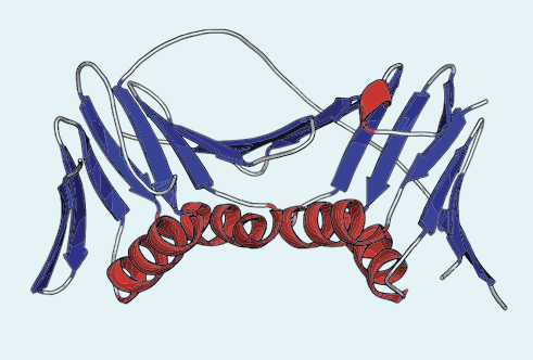
Tertiary structure
The tertiary structure is the functional three-dimensional shape of a polypeptide chain. This is the overall 3D structure that results from further folding of the secondary structures.
For a protein to be functional, it must at a minimum have a tertiary structure. The tertiary structure forms when secondary structures (alpha-helices, beta-pleated sheets, and random coils) fold further by forming various interactions and bonds between amino acids and R-groups in different sections of the polypeptide.
One important type of bond that stabilises tertiary structure is the disulphide bond, which is a strong covalent bond occurring between two sulphur atoms. Disulphide bonds often form between cysteine amino acids because these amino acids contain sulphur atoms in their R-groups. These bonds help stabilise the protein's three-dimensional structure.
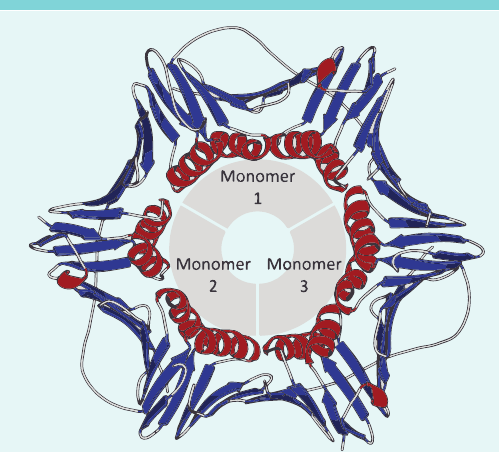
Quaternary structure
The quaternary structure is the level of protein structure where multiple polypeptide chains bond together, or other non-protein groups are added to form a fully functional protein.
Quaternary structure forms when two or more polypeptide chains, each with their own tertiary structure, join together. Polypeptide chains with tertiary or quaternary structure can also have a prosthetic group attached. A prosthetic group is a non-protein group bound to a protein, such as a vitamin or ion.
Not all proteins have a quaternary structure. Only proteins composed of multiple polypeptide chains, or those with prosthetic groups attached, are described as having quaternary structure.
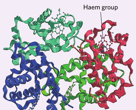
Haemoglobin: an example of quaternary structure
Haemoglobin is an excellent example of a protein with quaternary structure. This protein is responsible for carrying oxygen in red blood cells.
Worked Example: Haemoglobin Structure
Haemoglobin demonstrates quaternary structure in the following ways:
Multiple polypeptide chains: Haemoglobin is composed of four polypeptide chains bonded together.
Prosthetic groups: Within each of these chains, there is an iron ion embedded within a haem prosthetic group. The haem group is the non-protein component that actually binds to oxygen molecules, whilst the protein chains provide the structure necessary for this function.
This combination of multiple polypeptide chains and prosthetic groups makes haemoglobin a perfect example of quaternary structure.
The relationship between structure and function
The functional diversity of proteins arises from their ability to fold into an unlimited number of complex shapes and sizes. This diversity is possible because there are countless combinations of the 20 different amino acids that can be arranged in different sequences and lengths.
How primary structure determines folding
The folding of a protein into its functional tertiary or quaternary structure ultimately depends on its primary structure. The sequence of amino acids determines which R-groups are present and where they are located along the chain. These R-groups interact with each other in specific ways based on their chemical properties (such as whether they are hydrophobic or hydrophilic), forming different types of bonds that favour folding into particular three-dimensional structures.
The importance of correct amino acid sequence
Because protein folding depends on the primary structure, any changes to the original sequence of amino acids can have serious consequences. If the amino acid sequence is altered, the protein may no longer be able to fold correctly into its functional shape. This prevents the protein from functioning normally and can lead to disease.
The critical link between sequence and function:
Changes to the amino acid sequence can prevent correct protein folding, which in turn prevents normal protein function. This demonstrates why the primary structure is so fundamental - it determines everything that follows in protein structure and function.
For example, in muscle tissue, proteins serve as a core component of muscle fibres. By increasing protein intake through diet, you can support muscle growth and repair. However, proteins don't just contribute to muscle mass. As we've seen, they also perform numerous other essential functions including signalling and reception, transport, muscle contraction, storage, immunity, and structural support.
Remember!
Key Points to Remember:
-
Proteins are diverse biomacromolecules made of amino acid chains that perform many essential functions including acting as enzymes, transport proteins, structural proteins, defence proteins, motor proteins, storage proteins, receptors, and hormones.
-
Amino acids are the building blocks of proteins, each consisting of a central carbon bonded to a hydrogen atom, carboxyl group, amino group, and R-group. The R-group varies among 20 different amino acids and determines each amino acid's identity and properties.
-
Amino acids join via condensation reactions to form polypeptide chains, with peptide bonds linking adjacent amino acids. In this process, amino acids act as monomers that form protein polymers.
-
Proteins have four levels of structure: primary (amino acid sequence), secondary (alpha-helices, beta-pleated sheets, random coils), tertiary (functional 3D shape), and quaternary (multiple polypeptide chains bonded together). Not all proteins have quaternary structure.
-
The primary structure determines how a protein folds into its functional shape. Changes to the amino acid sequence can prevent correct folding and normal function, demonstrating the critical link between protein structure and function.