Structure of Skeletal Muscle (AQA A-Level Biology): Revision Notes
Structure of Skeletal Muscle
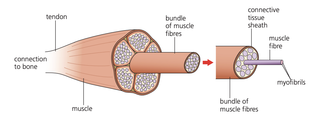
Overview of skeletal muscle organisation
Skeletal muscle forms the bulk of body muscle in vertebrates and attaches to bones, enabling voluntary movement under conscious control. These muscles function as effector organs that respond to nervous stimulation through contraction.
The structural organisation of skeletal muscle follows a hierarchical pattern, similar to how individual threads combine to form a strong rope. Just as rope threads are bundled into progressively larger units for maximum strength, muscle tissue consists of smaller components organised into increasingly larger structures.
This hierarchical organisation - from smallest to largest components - is fundamental to understanding how skeletal muscle generates such powerful forces. Each level of organisation contributes to the overall strength and coordination of muscle contraction.
At the most basic level, millions of tiny muscle fibres called myofibrils group together to form individual muscle fibres. These muscle fibres then bundle together to create the complete muscle structure. This arrangement maximises the force-generating capacity of the muscle.
Muscle fibre structure and composition
Individual muscle cells have undergone structural modifications to optimise their contractile function. Rather than existing as separate cells joined end-to-end (which would create weak junction points), muscle cells have fused together to form elongated muscle fibres.
The fusion of muscle cells into long fibres eliminates weak junction points that would occur between separate cells. This structural adaptation is crucial for generating continuous, powerful contractions along the entire length of the muscle.
These muscle fibres contain multiple nuclei and specialised cytoplasm called sarcoplasm, which surrounds the contractile elements. The sarcoplasm contains high concentrations of mitochondria and endoplasmic reticulum to support the energy demands of muscle contraction.
Within each muscle fibre, the contractile apparatus consists of numerous myofibrils - the actual contractile units responsible for generating force. Each myofibril contains the protein filaments that produce muscle contraction.
Protein filament composition
Myofibrils contain two main types of protein filament that interact to produce contraction:
Actin filaments (thin filaments) consist of two protein strands twisted around each other in a helical arrangement. These thinner filaments provide the framework for muscle contraction.
Myosin filaments (thick filaments) have a more complex structure, featuring long rod-shaped tails with bulbous heads projecting outward. These heads contain the molecular machinery necessary for generating contractile force.
The interaction between actin and myosin filaments forms the basis of the sliding filament theory of muscle contraction. The myosin heads act like molecular motors, pulling the actin filaments to generate force.
The arrangement of these filaments creates the characteristic appearance of skeletal muscle when viewed under a microscope.
Striped appearance and sarcomere structure
The regular arrangement of thick and thin filaments produces a distinctive striped pattern visible under light microscopy. This banding pattern results from the overlapping arrangement of actin and myosin filaments.
I bands (isotropic bands) appear lighter in colour because they contain only thin actin filaments. The centre of each I band contains a structure called the Z-line, which anchors the actin filaments.
A bands (anisotropic bands) appear darker because they contain both thick and thin filaments overlapping in this region. At the centre of each A band lies the H-zone, a lighter region containing only thick myosin filaments.
A sarcomere represents the functional unit of muscle contraction, defined as the distance between two adjacent Z-lines. When muscle contracts, sarcomeres shorten as the actin and myosin filaments slide past each other, changing the appearance of the banding pattern.
The sarcomere is like a repeating module of muscle contraction. Understanding its structure helps explain how the entire muscle shortens during contraction - thousands of sarcomeres working in unison create the overall muscle movement.
An additional protein called tropomyosin forms fibrous strands that wrap around actin filaments, playing a regulatory role in muscle contraction.
Types of muscle fibre
Skeletal muscles contain two distinct types of muscle fibre, with the proportion varying between different muscles and individuals:
Slow-twitch fibres contract more gradually and maintain contraction for extended periods, making them suitable for endurance activities like distance running. These fibres demonstrate several adaptations for sustained aerobic activity:
Slow-twitch fibre adaptations for endurance: Slow-twitch fibres are perfectly designed for sustained, aerobic exercise. Their adaptations work together to maintain steady energy production over long periods.
- Large stores of myoglobin (an oxygen-storing protein) give these fibres their characteristic red colour
- Rich blood vessel supply ensures adequate oxygen and glucose delivery
- Numerous mitochondria support continuous ATP production through aerobic respiration
Fast-twitch fibres generate rapid, powerful contractions but only for brief periods, making them ideal for explosive movements like weightlifting. These fibres show adaptations for anaerobic activity:
Fast-twitch fibre adaptations for power: Fast-twitch fibres sacrifice endurance for explosive power. Their adaptations enable rapid energy release for short bursts of intense activity.
- Higher concentrations of myosin filaments increase contractile force
- Elevated glycogen stores provide readily available energy
- High concentrations of enzymes supporting anaerobic respiration enable rapid ATP generation
- Phosphocreatine stores allow immediate ATP regeneration during intense activity
Key Points to Remember:
- Skeletal muscle has a hierarchical structure from whole muscle → muscle fibres → myofibrils → protein filaments
- Myofibrils contain actin (thin) and myosin (thick) filaments arranged in repeating units called sarcomeres
- The striped appearance results from overlapping filaments creating light I bands and dark A bands
- Slow-twitch fibres support endurance through aerobic adaptations, while fast-twitch fibres enable powerful short-term contractions
- Motor units consisting of a motor neurone and its associated muscle fibres allow precise control of muscle force