Ear as a sound detection system (AQA A-Level Physics): Revision Notes
📚 Revision Notes
10.2.1 Ear as a sound detection system
infoNote
The ear is a complex structure designed to detect sound, consisting of three main sections: the outer ear, middle ear, and inner ear. Each section plays a critical role in transmitting sound waves from the environment to the brain.
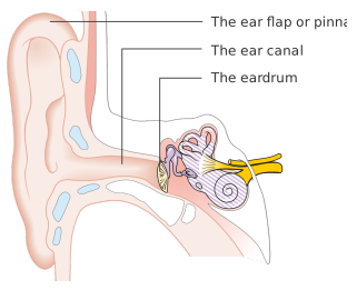
1. Outer Ear
- The outer ear is composed of the pinna (ear flap) and the external auditory canal.
- The pinna collects sound waves and, due to its shape, helps to determine the direction of the sound source. It directs these waves down the auditory canal towards the tympanic membrane (eardrum).
- The auditory canal contains wax glands, which produce wax to protect the eardrum by keeping it moist and flexible.
2. Middle Ear
- The middle ear consists of three small bones called the ossicles (malleus, incus, and stapes), as well as the Eustachian tube, oval window, and round window.
- Ossicles: The malleus (hammer) is connected to the eardrum, and the stapes (stirrup) is connected to the oval window. The ossicles work as a lever system to amplify sound vibrations. This amplification occurs with a factor of about 1.5.
- Eustachian Tube: This tube connects the middle ear to the throat, allowing it to remain at atmospheric pressure.
- Oval Window: This is a small membrane that vibrates in response to the stapes, creating pressure variations that are transferred into the inner ear.
- Round Window: This structure provides flexibility in the inner ear, allowing fluid movement necessary for sound transmission. Through the lever action of the ossicles, the pressure at the eardrum is amplified by a factor of 20 by the time it reaches the oval window.
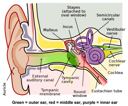
3. Inner Ear
- The inner ear is filled with a fluid called perilymph that facilitates sound transmission. It includes the cochlea, auditory nerve, and balance organs (semicircular canals).
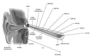
- Cochlea: Shaped like a spiral, the cochlea is filled with a different fluid called endolymph. Vibrations pass along the length of the cochlea, causing the basilar membrane to vibrate.
- Hair Cells: The basilar membrane contains hair cells that generate electrical signals in response to vibrations, which are then sent to the brain through the auditory nerve.
- Frequency Detection: Different regions of the basilar membrane respond to specific frequencies. High-frequency sounds activate hair cells near the base of the cochlea, while low-frequency sounds affect hair cells near the apex. As sound waves move through each section of the ear, the amplitude of the sound decreases, but the frequency remains constant.
infoNote
Key Points of Sound Transmission in the Ear
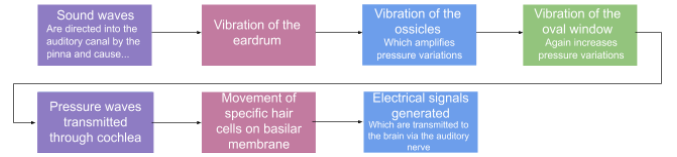
- Sound waves are collected by the pinna and directed into the auditory canal.
- The tympanic membrane vibrates in response to these waves.
- The ossicles amplify and transmit these vibrations to the oval window.
- Vibrations at the oval window create pressure waves in the cochlear fluid.
- These waves stimulate hair cells on the basilar membrane.
- Electrical signals generated by the hair cells are transmitted to the brain via the auditory nerve, allowing us to perceive sound.