Image detection and enhancement (AQA A-Level Physics): Revision Notes
10.5.2 Image detection and enhancement
X-ray Detection and Digital Imaging:
When using traditional photographic film for X-ray imaging, only around 0.1% of the X-ray energy is absorbed, which means the exposure time must be extended to produce a clear image. This increases the radiation dose to the patient and raises the risk of image blurring if the patient moves.
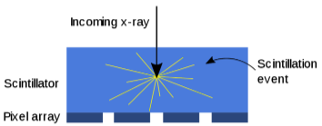
To improve efficiency, Flat Panel (FTP) Detectors are now commonly used. These detectors include a scintillator, which is a material that emits light photons when it absorbs high-energy X-rays. The intensity of the light emitted by the scintillator is directly proportional to the X-ray energy absorbed. Photodiodes, which are sensitive to this light, convert it into electronic signals, forming a digital image.
Each photodiode acts as an individual pixel in the image, with the total number of photodiodes determining the resolution. The digital data is scanned, converted, and processed, allowing for high-resolution digital X-ray images.
Advantages of FTP Detectors Over Film:
- Lower Radiation Dose: FTP detectors are more sensitive, allowing a shorter exposure time and reducing patient radiation exposure.
- Instant Digital Images: The images can be instantly accessed, shared, and evaluated.
- Higher Resolution: FTP images have better resolution and less distortion than film-based images.
- Portability: FTP detectors are compact and lightweight, making them easier to transport around hospitals.
Contrast in X-ray Imaging:
A contrast medium can be used to increase the clarity of X-ray images, especially when there's little difference between the proton numbers of the tissues being examined. For example, a barium meal is used as a contrast agent for gastrointestinal imaging. This involves:
- The patient swallowing a barium mixture.
- The barium highlights the gastrointestinal tract by creating a stark contrast with surrounding tissues.
Enhancement Techniques in X-ray Imaging:
- X-ray Opaque Contrast Medium: These media absorb more X-rays, showing as white areas on X-ray film.
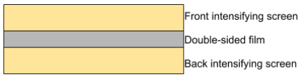
- Intensifying Screens: Made of fluorescent crystals that emit visible light when exposed to X-rays. This screen is placed next to the film and reduces the exposure time required, lowering patient radiation.
- Film Placement and Orientation: Film is placed between intensifying screens to maximise light capture, and reduce the radiation required.
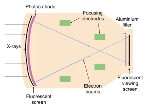
Fluoroscopic Image Intensification (for live X-ray imaging):
This method uses a fluorescent screen instead of film, which emits a dim light image when exposed to X-rays. Image intensifiers magnify this light by accelerating electrons through a potential difference to increase brightness:
- Electrons are emitted from a photocathode and accelerated to hit a fluorescent viewing screen, creating a brighter image.
- Focusing electrodes ensure the image remains sharp and reduce patient dose by enabling low-intensity X-rays.
To manage brightness effectively:
- The fluorescent screen closest to the photocathode has an aluminium layer that blocks emitted light from scattering back, improving image contrast. This method provides real-time X-ray imaging but requires a higher radiation dose, making it suitable for specific diagnostics only.