Interpreting NMR Spectra (OCR A-Level Chemistry A): Revision Notes
Interpreting NMR Spectra
Introduction to interpreting proton NMR spectra
Nuclear magnetic resonance (NMR) spectroscopy is a powerful analytical technique used to identify organic compounds. When interpreting proton (H) NMR spectra, there is no single rigid procedure you must follow - the approach can be flexible depending on which features stand out most clearly in the spectrum. However, using a systematic step-by-step method will help ensure you don't miss important information.
While you can approach NMR interpretation flexibly, following a systematic method ensures you analyze all available information and don't overlook crucial details that could lead to incorrect structure assignments.
The key pieces of information available from a proton NMR spectrum are:
- Peak positions (chemical shift, , measured in ppm) - tell you about the chemical environment of protons
- Peak areas (integration values) - tell you the relative numbers of protons in each environment
- Splitting patterns - tell you about adjacent protons on neighbouring carbon atoms
By combining these three types of information with chemical shift data tables and your knowledge of organic structures, you can deduce the structure of unknown compounds.
Systematic approach to interpreting proton NMR
A reliable four-step approach involves:
Step 1: Analyse the types of proton present and how many of each type
Start by counting how many distinct peaks appear in the spectrum - this tells you how many different proton environments exist in the molecule. Then examine the relative peak areas (integration values) to determine the ratio of protons in each environment.
The integration values are proportional to the number of protons producing each signal. For example, if you see integration values of 3:2:3, this means the ratio of protons in those three environments is 3:2:3. You may need to simplify ratios or recognise that they should match the molecular formula.
From the integration ratio, you can deduce what types of groups might be present. For instance:
- An integration of 3 often suggests a group
- An integration of 2 often suggests a group
- An integration of 1 suggests a single proton
Step 2: Analyse the splitting patterns to find information about adjacent protons
The splitting pattern of each peak provides crucial information about adjacent protons through the n + 1 rule:
where is the number of hydrogen atoms on adjacent carbon atoms.
The n + 1 rule is fundamental to NMR interpretation
Always remember: the number of peaks you see in a splitting pattern equals the number of adjacent protons plus one. This relationship allows you to determine the structure of neighbouring groups even without seeing them directly.
Common splitting patterns include:
- Singlet: No adjacent protons (, so peak)
- Doublet: 1 adjacent proton (, so peaks)
- Triplet: 2 adjacent protons (, so peaks)
- Quartet: 3 adjacent protons (, so peaks)
- Quintet: 4 adjacent protons (, so peaks)
- Heptet: 6 adjacent protons (, so peaks)
By identifying splitting patterns, you can work out which groups are adjacent to each other in the molecule. For example, a triplet-quartet combination strongly suggests an ethyl ester group (), where:
- The appears as a triplet (split by 2 adjacent H atoms on )
- The appears as a quartet (split by 3 adjacent H atoms on )
Recognizing Common Patterns
The triplet-quartet combination is one of the most characteristic patterns in proton NMR. Once you recognize this pattern, you can immediately identify an ethyl group () in the structure. This is a powerful shortcut in structure determination!
Step 3: Analyse the chemical shifts for the types of proton
Each type of proton appears at a characteristic chemical shift value depending on its chemical environment. Protons closer to electronegative atoms or groups (like oxygen, nitrogen, or carbonyl groups) appear at higher chemical shift values (further downfield, towards the left of the spectrum) because they are more deshielded.
You should use chemical shift data tables to identify which functional groups are present. Common ranges include:
- ppm: protons on carbons bonded to other carbons ()
- ppm: protons on carbons adjacent to groups
- ppm: protons on carbons bonded to oxygen ()
- ppm: aldehyde protons ()
- ppm: carboxylic acid protons ()
Understanding Deshielding
Electronegative atoms and groups pull electron density away from nearby protons, reducing the shielding effect of surrounding electrons. This makes these protons more exposed to the applied magnetic field, causing them to resonate at higher frequencies and appear further downfield (higher values) in the spectrum.
Step 4: Combine the information to suggest a structure
Using all the evidence from steps 1-3, piece together the molecular structure. Check that your proposed structure:
- Has the correct molecular formula
- Has the right number of proton environments
- Shows the observed splitting patterns
- Has protons in chemical environments matching the observed chemical shifts
Validating Your Structure
Your proposed structure must satisfy ALL the spectroscopic evidence. If even one piece of data doesn't fit, your structure is incorrect. Always systematically check each requirement before finalizing your answer.
If your structure doesn't fit all the evidence, revisit your interpretation of the spectrum.
Worked example 1: Interpreting the spectrum of an isomer of C₄H₈O₂
Worked Example: Complete Interpretation of a Proton NMR Spectrum
Let's work through a complete interpretation of a proton NMR spectrum for a compound with molecular formula .
Step 1: Analyse the types of proton present
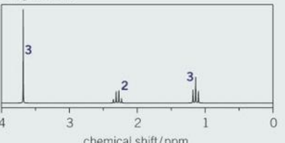
The spectrum shows three distinct peaks, indicating three different proton environments. Looking at the relative peak areas (integration values), the ratio is 3:2:3.
This ratio suggests:
- A peak at ppm representing 3 protons (likely )
- A peak at ppm representing 2 protons (likely )
- A peak at ppm representing 3 protons (likely )
Step 2: Analyse the splitting patterns
Examining each peak more carefully:
- The peak at ppm is a singlet, which tells us there are no adjacent protons (by the rule: )
- The peak at ppm is a triplet, indicating an adjacent group (by the rule: )
- The peak at ppm is a quartet, indicating an adjacent group (by the rule: )
The combination of a triplet at ppm and a quartet at ppm is characteristic of an ethyl group ().
Step 3: Analyse the chemical shifts
Using chemical shift data:
- The peak at ppm indicates protons in the environment , suggesting a sequence like
- The peak at ppm indicates protons in the environment , suggesting a sequence like
- The peak at ppm indicates protons in the environment , suggesting a sequence like
Step 4: Combine the information
Putting together all the evidence:
- We have a group (from the triplet-quartet combination)
- We have a group (from the chemical shift at 2.3 ppm)
- We have a group (from the singlet at 3.7 ppm)
- The molecular formula is , suggesting an ester
The structure must be methyl propanoate:
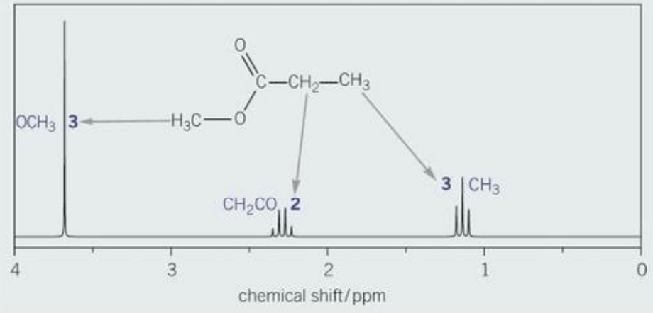
This annotated spectrum shows how each group of protons corresponds to its peak, confirming our interpretation.
Worked example 2: Interpreting the spectrum of an isomer of C₃H₇Cl
Worked Example: Identifying Structure from Heptet-Doublet Pattern
Now let's interpret a proton NMR spectrum for a compound with molecular formula .

Step 1: Analyse the types of proton present
This spectrum shows two peaks, so there are two different proton environments. The integration ratio is 1:6.
This suggests:
- A peak at ppm representing 1 proton ()
- A peak at ppm representing 6 protons ()
Step 2: Analyse the splitting patterns
Looking at the splitting:
- The peak at ppm is a heptet (7 peaks), which indicates 6 adjacent protons (by the rule: ). This can only arise from two adjacent groups:
- The peak at ppm is a doublet, indicating 1 adjacent proton (by the rule: )
The combination of a doublet at ppm and a heptet at ppm tells us the structure contains .
Step 3: Analyse the chemical shifts
Using chemical shift data:
- The peak at ppm indicates the environment , supporting a sequence
- The peak at ppm indicates the environment , supporting the sequence
Step 4: Combine the information
Combining all the evidence from steps 1-3:
- We have two equivalent groups
- We have one group bonded to chlorine
- The is adjacent to both groups
The structure must be 2-chloropropane:
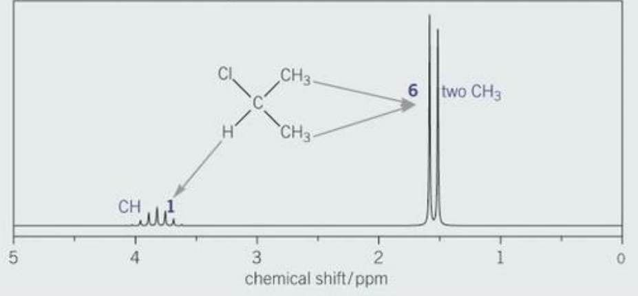
This annotated spectrum confirms our deduced structure, showing how the two methyl groups produce the large peak at 1.6 ppm and the single CH group produces the smaller, split peak at 3.8 ppm.
Predicting NMR spectra
While interpreting NMR spectra involves working from the spectrum to deduce the structure, you also need to be able to work in the opposite direction - predicting what the NMR spectrum will look like for a given structure. This skill is essential for confirming proposed structures and understanding spectroscopic data.
To predict an NMR spectrum, you reverse the interpretation process:
- Draw out the structure clearly
- Identify different chemical environments
- Predict chemical shift values for each environment
- Predict relative peak heights from the number of protons
- Predict splitting patterns using the rule
You can predict both carbon-13 (C) NMR and proton (H) NMR spectra using this approach.
Worked example 3: Predicting a carbon-13 NMR spectrum
Worked Example: Predicting a Carbon-13 NMR Spectrum
Let's predict the carbon-13 NMR spectrum for 2-hydroxypropanoic acid (lactic acid), .
Step 1: Draw out the structure
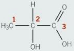
The structure shows the skeletal formula of lactic acid with three carbon atoms.
Step 2: Identify the number of chemical environments
Looking at the carbon atoms in the structure:
- Carbon 1: group bonded to C-OH
- Carbon 2: group bonded to OH and to the rest of the chain
- Carbon 3: carboxylic acid carbon with C=O
These three carbon atoms are in different chemical environments, so we expect three peaks in the carbon-13 NMR spectrum.
Step 3: Predict the chemical shifts
Using carbon-13 chemical shift data:
- Carbon-1 is of type , so we predict a peak at ppm
- Carbon-2 is of type , so we predict a peak at ppm
- Carbon-3 is of type (carboxylic acid), so we predict a peak at ppm
Checking the prediction
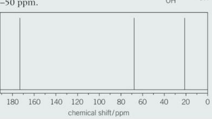
The actual carbon-13 NMR spectrum shows three peaks at approximately 20 ppm, 80 ppm, and 170 ppm, which matches our predictions very well. The peaks appear in the expected regions for the three different carbon environments.
Worked example 4: Predicting a proton NMR spectrum
Worked Example: Predicting a Proton NMR Spectrum with Splitting Patterns
Now let's predict the proton NMR spectrum for the same compound, 2-hydroxypropanoic acid, .
Step 1: Draw the structure and identify the number of chemical environments
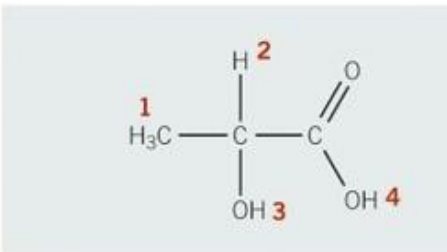
This numbered structure shows four different types of protons, so we expect four peaks in the proton NMR spectrum:
- Protons 1: group
- Proton 2: group
- Proton 3: group on carbon 2
- Proton 4: group
Step 2: Predict the chemical shifts
Using proton chemical shift data:
- Protons 1 () are of type , giving a peak at ppm
- Proton 2 () is of type , giving a peak at ppm
- Proton 3 () can appear anywhere in the range ppm (broad, variable)
- Proton 4 () is of type carboxylic acid, giving a peak at ppm
Step 3: Predict the relative peak heights
The relative peak heights depend on the number of each type of proton. For protons 1-4, the ratio would be 3:1:1:1 ().
Step 4: Predict the splitting patterns
Using the rule:
- Protons 1 () have one adjacent proton (the ), so they appear as a doublet ()
- Proton 2 () has three adjacent protons (the group), so it appears as a quartet ()
- Protons 3 and 4 ( and ) typically give broad peaks without clear splitting due to rapid proton exchange
Checking the prediction
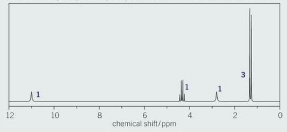
The actual proton NMR spectrum shows the predicted features:
- A peak with integration 3 around ppm for the group
- Peaks around ppm for the group
- A peak around ppm for the proton
Notice that the quartet for the group at ppm appears at the upper end of the predicted range (3.0-4.2 ppm). This is because this group is adjacent to both an oxygen atom (from the ) and a carbonyl group (from ), both of which cause additional deshielding and shift the signal further downfield.
Key Points to Remember:
- Use a systematic approach: Analyse peak numbers and areas, splitting patterns, and chemical shifts in a logical order
- The n + 1 rule is crucial: Number of peaks in splitting pattern = , where is the number of adjacent protons
- Integration ratios show relative numbers of protons: Use these to identify groups like , , etc.
- Chemical shift values indicate functional groups: Higher values (downfield) indicate protons near electronegative atoms or groups
- Triplet-quartet combinations suggest ethyl groups: Look for characteristic 3:2 ratios
- You can predict spectra from structures: Work through the same steps in reverse - identify environments, predict shifts, and determine splitting patterns
- Singlets indicate isolated groups: No adjacent protons means no splitting
- Carboxylic acid and alcohol protons give broad peaks: Due to proton exchange, these often don't show clear splitting
Exam focus checklist
✓ Can you identify the number of proton environments from a spectrum?
✓ Can you calculate the ratio of protons from integration values?
✓ Can you apply the rule to interpret splitting patterns?
✓ Can you identify common patterns like triplet-quartet combinations?
✓ Can you use chemical shift data to identify functional groups?
✓ Can you combine all spectroscopic evidence to propose a structure?
✓ Can you predict a carbon-13 NMR spectrum from a given structure?
✓ Can you predict a proton NMR spectrum including chemical shifts, integrations, and splitting patterns?
✓ Do you know typical chemical shift ranges for common functional groups?
✓ Can you explain why certain groups cause deshielding and higher chemical shifts?