NMR Spectroscopy (OCR A-Level Chemistry A): Revision Notes
NMR Spectroscopy
Introduction to NMR spectroscopy
Nuclear magnetic resonance (NMR) spectroscopy is one of the most powerful analytical techniques available to chemists. Developed approximately 70 years ago, this method has completely transformed how we analyze and understand the structure of organic compounds. The technique works by combining a very powerful magnetic field with radio frequency (RF) radiation to probe the atomic nuclei within molecules.
When the correct combination of magnetic field strength and frequency is applied, certain atomic nuclei can absorb radiation. The energy absorbed during this process is measured and displayed as an NMR spectrum, which provides detailed information about the molecular structure without destroying the sample.
One of the key advantages of NMR spectroscopy is that it is non-destructive – the sample can be recovered after analysis by simply evaporating the solvent. This makes it invaluable for analyzing precious or limited samples.
What is nuclear magnetic resonance?
Nuclear spin
To understand NMR spectroscopy, you need to know about a property called nuclear spin. Just as electrons possess a property called spin, some atomic nuclei also have this characteristic. The key rule is that a nucleus has spin if it contains an odd number of nucleons (protons and neutrons combined).
For organic chemists, this is particularly important because most organic molecules contain carbon and hydrogen atoms. The isotopes H and C are the most abundant forms in organic compounds. However, C has an even number of nucleons and therefore shows no NMR signal. Instead, we rely on:
- H (hydrogen-1): The most common isotope, consisting of just one proton. This is by far the most frequently used nucleus for NMR analysis.
- C (carbon-13): Makes up only about 1.1% of carbon atoms but has an odd number of nucleons and can be detected.
Because H consists of a single proton, H NMR spectroscopy is commonly referred to as proton NMR. While NMR can detect other isotopes with odd nucleon numbers such as F and P, H and C NMR are the most commonly used forms of this analysis.
The resonance phenomenon
The term "resonance" in NMR refers to what happens when nuclei absorb energy. Both electrons and nuclei can exist in different spin states, each with a different energy level. For a nucleus, there are two possible spin states with distinct energies.
When you apply the right combination of a strong magnetic field and radio frequency radiation, something remarkable happens: the nucleus can absorb energy and rapidly flip between these two spin states. This process is called resonance, and it's this behaviour that gives nuclear magnetic resonance its name.
The two spin states of a nucleus have slightly different energy levels. When nuclei absorb the exact amount of energy matching this energy difference, they can flip from the lower energy state to the higher energy state – this is the resonance phenomenon that NMR spectroscopy detects.
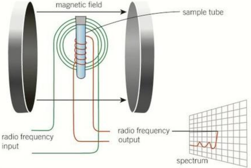
The NMR spectrometer
Equipment and magnetic fields
NMR spectroscopy requires specialized and sophisticated equipment. The radio frequency radiation used in NMR has considerably less energy than the infrared radiation used in IR spectroscopy. Because of this lower energy, the frequency needed for resonance is directly proportional to the strength of the applied magnetic field.
For resonance to occur, you need:
- A very strong magnetic field that is completely uniform throughout
- A constant radio frequency matching the magnetic field strength
To achieve these conditions, NMR spectrometers use super-conducting electromagnets that must be cooled to extremely low temperatures (typically 4 K) using liquid helium. This creates the powerful and stable magnetic field necessary for accurate measurements.
The extreme conditions required for NMR spectroscopy – including temperatures near absolute zero (4 K or -269°C) and magnetic fields tens of thousands of times stronger than Earth's magnetic field – explain why NMR spectrometers are both expensive and require specialized facilities to operate.
Modern routine NMR spectrometers typically operate at radio frequencies of 100, 200, or 400 MHz. University research facilities often have these instruments, but they are far too expensive for most schools and colleges to own.
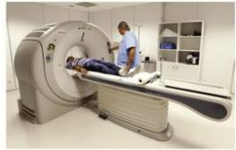
Applications beyond chemistry
The same technology used in NMR spectrometers also powers the MRI (magnetic resonance imaging) body scanners found in hospitals. These medical devices use identical principles to create detailed images of the inside of the human body, demonstrating the wide-ranging applications of this technology.
Chemical shift and TMS
Understanding chemical shift
In an organic molecule, every carbon and hydrogen atom is bonded to other atoms. Each atom is surrounded by electrons, which create their own small magnetic fields. These electron clouds shield the nucleus from the external magnetic field applied by the spectrometer.
The presence of these surrounding electrons means that the actual magnetic field experienced by a nucleus is slightly different from the applied field. This shielding effect alters the energy and radio frequency required for resonance to occur. The variation in frequency needed is measured on a scale called chemical shift, represented by the symbol δ (delta) and measured in parts per million (ppm).
Tetramethylsilane (TMS) as the reference standard
To make chemical shift measurements meaningful and comparable, we need a reference point. The standard reference compound used is tetramethylsilane, abbreviated as TMS, with the formula .
TMS is chosen as the standard because:
- It produces a single, sharp peak in the spectrum
- It is chemically inert and doesn't react with samples
- All its hydrogen atoms are equivalent
- Its peak appears away from most organic compound signals
By definition, TMS is assigned a chemical shift value of 0 ppm. All other chemical shifts in a spectrum are measured relative to this standard.
Factors affecting chemical shift
The amount of chemical shift experienced by a nucleus depends primarily on its chemical environment – particularly the presence of nearby electronegative atoms. Different types of bonds and molecular environments produce characteristic chemical shift values.
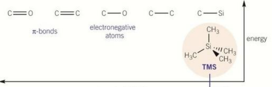
The diagram shows how different bonding situations affect chemical shift:
- C=O π-bonds and C=C double bonds cause larger chemical shifts (appear further left on the spectrum)
- C-O bonds with electronegative oxygen atoms cause moderate shifts
- C-C single bonds cause smaller shifts
- C-Si bonds in TMS cause the smallest shift (0 ppm)
The more electronegative the nearby atoms, the more the nucleus is deshielded and the larger the chemical shift value. This creates a predictable pattern that allows chemists to identify different functional groups in a molecule.
Important exam tip: In an NMR spectrum, chemical shift values increase from right to left. The TMS reference peak at 0 ppm always appears at the right-hand end of the scale, and chemical shift values increase as you move left across the spectrum.
Mapping molecular structure
The beauty of NMR spectroscopy is that because different chemical environments produce absorption peaks at characteristic chemical shifts, you can map out the arrangement of carbon and hydrogen atoms in a molecule. This analysis can be performed without needing to carry out traditional chemical tests and without destroying the organic compound being studied.
Running the spectrum
Sample preparation
To obtain an NMR spectrum, you must first prepare your sample correctly:
- Dissolve the sample in an appropriate solvent
- Add a small amount of TMS as the reference standard
- Place the solution in a narrow NMR sample tube – these are specialized tubes designed to fit inside the spectrometer
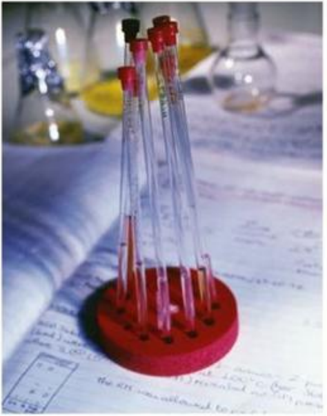
The NMR sample tube is a precision-made piece of glassware, carefully designed to have uniform wall thickness and to fit exactly within the spectrometer's magnetic field. The sample solution typically needs to be only a few milliliters in volume.
The measurement process
Once the sample tube is prepared:
- The tube is placed inside the NMR spectrometer between the poles of the powerful electromagnet
- The tube is spun rapidly to ensure the magnetic field is experienced uniformly throughout the sample and to eliminate any imperfections
- The spectrometer is zeroed against the TMS standard
- The sample is given a pulse of radiation containing a range of radio frequencies
- The spectrometer maintains a constant magnetic field throughout the measurement
Any absorptions of energy resulting from resonance are detected by the instrument and displayed on a computer screen as a spectrum. Once analysis is complete, the sample can be recovered by evaporating the solvent, meaning the compound is not destroyed during analysis.
Deuterated solvents
Why use deuterated solvents?
Most organic molecules contain both carbon and hydrogen atoms. When you run an NMR spectrum, these atoms will naturally produce signals in both C and H NMR spectra. However, if you dissolved your sample in a normal solvent (which also contains hydrogen atoms), the hydrogen atoms from the solvent would also produce signals, potentially obscuring or interfering with the signals from your compound of interest.
To avoid this problem, chemists use deuterated solvents. In these solvents, the H atoms have been replaced by H atoms (deuterium, symbol D). Deuterium has an even number of nucleons (one proton and one neutron), so it produces no signal in the frequency ranges used for H and C NMR spectroscopy.
Deuterium (H or D) is invisible to both H and C NMR spectroscopy because it has an even number of nucleons (1 proton + 1 neutron = 2 nucleons). This makes deuterated solvents essential for preventing interference from solvent hydrogen atoms in the spectrum.
Common deuterated solvents
The most commonly used deuterated solvent in NMR spectroscopy is deuterated trichloromethane, (also called deuterated chloroform). However, there's one complication: even though the hydrogen has been replaced with deuterium, the carbon atoms are still present. This means will still produce a peak in a carbon-13 NMR spectrum.
Fortunately, modern NMR spectrometers can handle this issue. The computer controlling the instrument usually filters out the solvent peak before displaying the spectrum, ensuring you only see peaks from your compound of interest.
Remember!
Key Points to Remember:
- Nuclear spin exists in nuclei with an odd number of nucleons; the most important for organic chemistry are H (proton NMR) and C
- Resonance occurs when nuclei absorb energy and flip between different spin states in the presence of a strong magnetic field and radio frequency radiation
- TMS (tetramethylsilane, ) is the universal reference standard for chemical shift measurements and is assigned a value of 0 ppm
- Chemical shift (δ) measures the variation in resonance frequency caused by the chemical environment, particularly the shielding effect of electrons and the presence of electronegative atoms
- Deuterated solvents like are essential to prevent solvent hydrogen atoms from interfering with the spectrum
- Chemical shift increases from right to left on an NMR spectrum, with TMS at the right-hand end (0 ppm)
- NMR is a non-destructive technique – the sample can be recovered after analysis
Exam focus checklist:
✓ Be able to explain what nuclear spin is and why H and C are important
✓ Understand the role of TMS as the reference standard at 0 ppm
✓ Know that chemical shift is affected by the chemical environment, especially electronegative atoms
✓ Remember that deuterated solvents prevent interference from solvent hydrogen
✓ Understand that NMR requires a strong magnetic field, radio frequency radiation, and a spinning sample
✓ Be able to explain why the sample is dissolved in a deuterated solvent with TMS added