Proteins (AQA A-Level Biology): Revision Notes
Structure of Proteins
Unlike carbohydrates and lipids, which show relatively little variation between species, proteins are incredibly diverse. Each organism produces thousands of different proteins, making them vital for structure and function in biological systems.
Despite having only 20 naturally occurring amino acids as building blocks, proteins can form an almost infinite variety of structures and functions. This diversity arises from the different ways these amino acids can be arranged and combined.
Primary structure: polypeptide formation
Peptide bond formation
Amino acids link together through peptide bonds formed by condensation reactions. During this process:
- The carboxyl group of one amino acid combines with the amino group of another
- A water molecule is eliminated
- A new covalent bond forms between the carbon of the first amino acid and the nitrogen of the second
This peptide bond connects the two amino acids, creating a dipeptide. The process can be reversed through hydrolysis, where water is added to break the peptide bond.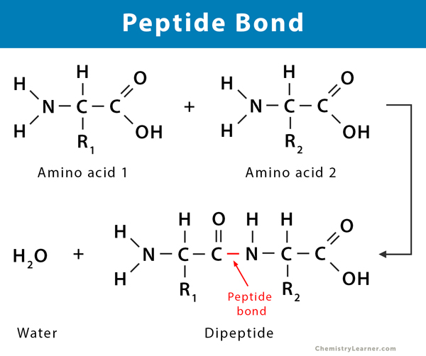
Worked Example: Peptide Bond Formation
When two amino acids join together:
- Amino acid 1:
- Amino acid 2:
The condensation reaction produces:
- Dipeptide:
- Water molecule eliminated:
The new peptide bond is the linkage between the two amino acids.
Polymerisation to form polypeptides
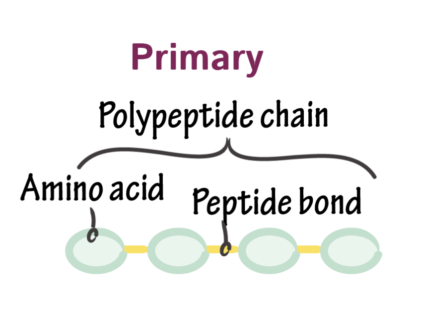
Through repeated condensation reactions, many amino acids can join together in a process called polymerisation. The resulting chain of hundreds of amino acids forms a polypeptide.
The primary structure of a protein refers to the specific sequence of amino acids in its polypeptide chain(s). This sequence is determined by DNA and is crucial because it determines all higher levels of protein structure and, consequently, the protein's function.
Secondary structure
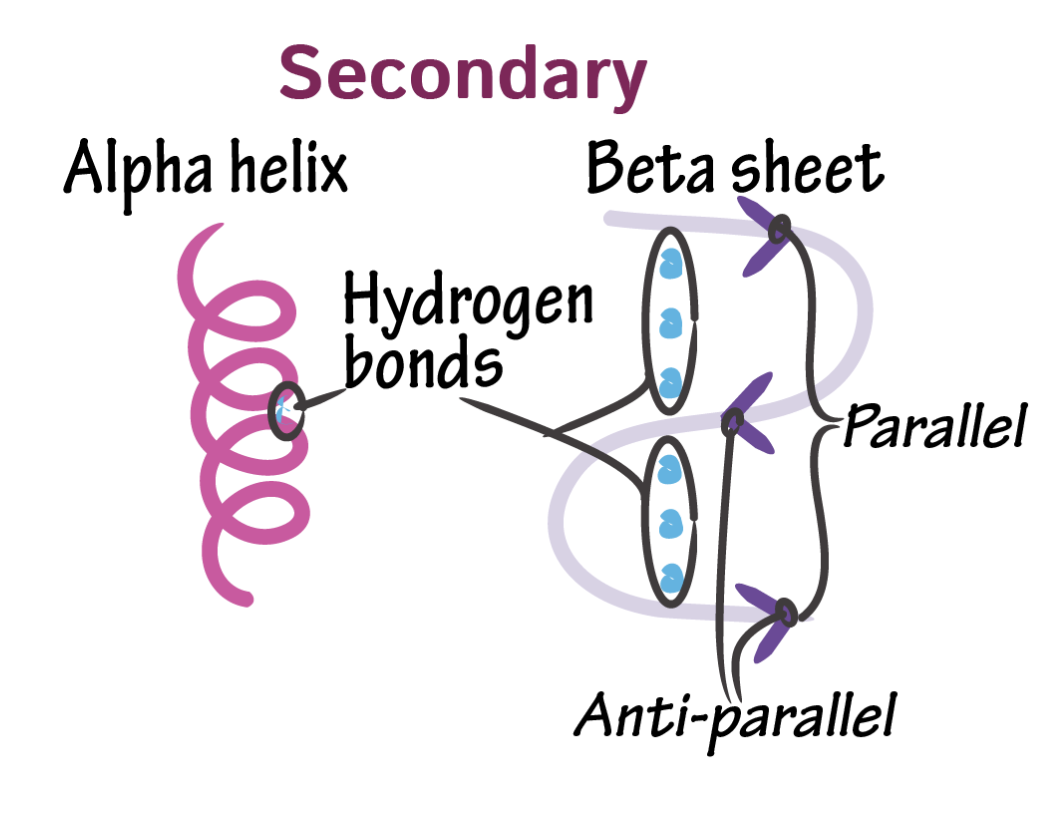
The secondary structure describes the shape that polypeptide chains adopt due to hydrogen bonding between different parts of the chain.
Hydrogen bond formation
Within polypeptide chains, the backbone contains repeating and groups from the amino and carboxyl components of amino acids. The hydrogen in groups carries a slight positive charge, while the oxygen in groups carries a slight negative charge. These opposite charges allow hydrogen bonds to form between different parts of the chain.
Hydrogen bonds are individually weak compared to covalent bonds, but when many hydrogen bonds form along a polypeptide chain, they provide significant stability to the protein's secondary structure.
The α-helix
The most common secondary structure is the α-helix (alpha helix), where hydrogen bonding causes the polypeptide chain to twist into a regular spiral shape. This coiled structure is maintained by hydrogen bonds between amino acids that are four positions apart in the sequence.
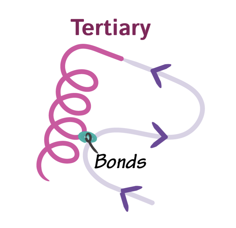
Tertiary structure
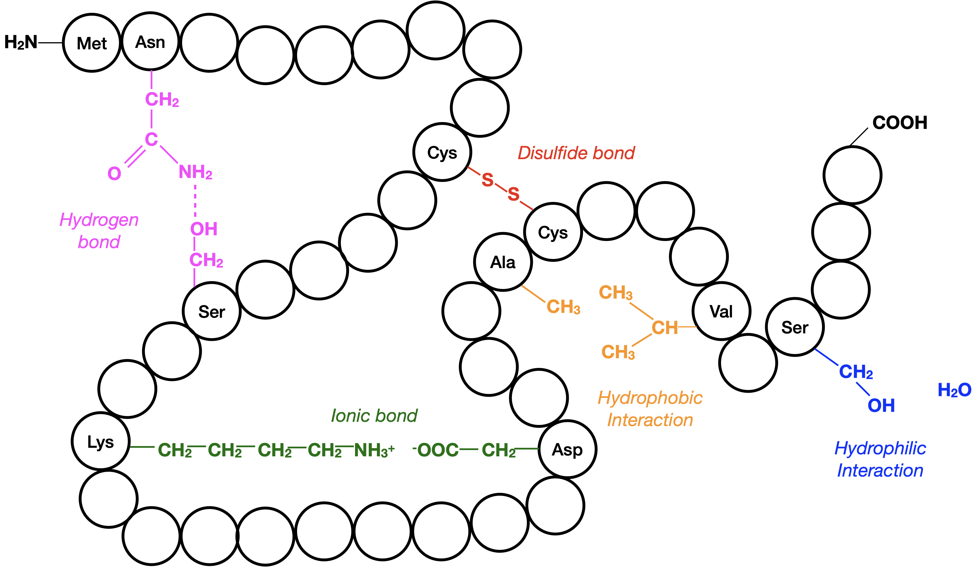
Tertiary structure refers to the complex three-dimensional shape that results from further folding and twisting of the secondary structure. This level of organisation is maintained by several types of chemical bonds:
Types of bonds
- Disulphide bridges: Strong covalent bonds that form between cysteine amino acids containing sulphur. These bonds are relatively stable and not easily broken
- Ionic bonds: Form between amino acids with oppositely charged R groups. These bonds are weaker than disulphide bridges and can be disrupted by pH changes
- Hydrogen bonds: Additional hydrogen bonds form between various R groups throughout the structure. Though individually weak, collectively they provide significant stability
Importance of tertiary structure
The specific three-dimensional shape of a protein is critical for its function. This shape allows the protein to:
- Recognise and bind to specific molecules
- Interact with other proteins in precise ways
- Carry out its biological role effectively
Even a single amino acid change in the primary structure can alter the tertiary structure, potentially destroying the protein's function.
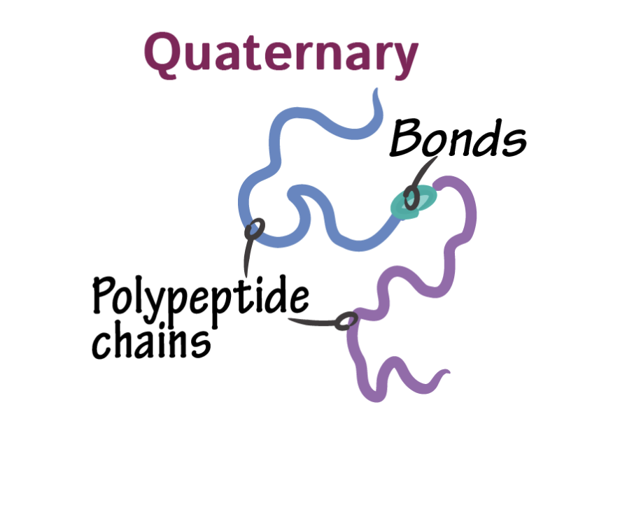
Quaternary structure
Many proteins consist of multiple polypeptide chains that associate together to form the final functional protein. This quaternary structure involves:
Multiple polypeptide chains
Large, complex proteins often contain several separate polypeptide chains that fit together like pieces of a puzzle. Each chain contributes to the overall structure and function of the protein.
Prosthetic groups
Some proteins also include prosthetic groups - non-protein components that are essential for function. For example, haemoglobin contains iron-containing haem groups that are crucial for oxygen binding.
The quaternary structure emphasises that while the primary structure (amino acid sequence) determines the initial folding, the final three-dimensional arrangement of multiple components creates the functional protein.
Key Points to Remember:
- Amino acids are the monomers that combine via peptide bonds to form polypeptide polymers
- Primary structure (amino acid sequence) determines all higher levels of protein organisation
- Secondary structure results from hydrogen bonding creating shapes like the α-helix
- Tertiary structure involves complex 3-D folding maintained by disulphide bridges, ionic bonds, and hydrogen bonds
- The biuret test detects proteins by producing a purple colour when peptide bonds are present