The Nerve Impulse (AQA A-Level Biology): Revision Notes
The Nerve Impulse
What is a nerve impulse?
A nerve impulse is a self-propagating wave of electrical activity that travels along the axon membrane. It represents a temporary reversal of the electrical potential difference across the axon membrane, switching between two main states: the resting potential and the action potential.
Resting potential
Establishing the resting potential
The resting potential is the electrical charge difference across an axon membrane when the neurone is not transmitting an impulse. In humans, this typically measures -65mV, though it can range from -50 to -90mV depending on the cell type.
The resting potential is established and maintained through three key mechanisms: membrane structure and permeability, active transport mechanisms, and specific ion distribution patterns.
The resting potential is established and maintained through several key mechanisms:
Membrane structure and permeability
- The phospholipid bilayer of the axon plasma membrane prevents free movement of sodium and potassium ions across it
- Channel proteins span this phospholipid layer, containing ion channels that allow specific ions to pass through
- Some channels have 'gates' that can open or close to control ion movement through facilitated diffusion
- Different voltage-gated channels exist for sodium and potassium ions
Active transport mechanisms
- Carrier proteins actively transport ions against their concentration gradients
- The sodium-potassium pump is crucial - it actively transports sodium ions out of the axon and potassium ions into the axon
- This pump moves three sodium ions out for every two potassium ions that move in, contributing to the negative charge inside
Ion distribution at rest
- Most potassium ion channels remain open, allowing potassium to diffuse out of the axon
- Most sodium ion channels are closed, preventing sodium influx
- More sodium ions accumulate in the tissue fluid outside the axon
- More potassium ions remain in the cytoplasm than in the tissue fluid
- This creates an electrochemical gradient with the inside of the axon negative relative to the outside
Action potential
Triggering an action potential
When a stimulus of sufficient size is detected by a receptor, it causes a temporary reversal of charges on either side of the axon membrane. If the stimulus is large enough, the negative charge of approximately -65mV inside the membrane becomes a positive charge of around +40mV. This is the action potential, and the membrane is said to be depolarised.
Stages of action potential
The action potential occurs through six distinct stages, involving voltage-gated channels that open or close depending on the voltage across the membrane:
Stage 1 - Resting state
- Some potassium voltage-gated channels are open (permanently open ones)
- Sodium voltage-gated channels are closed
- Membrane potential maintained at -65mV
Stage 2 - Stimulus and initial depolarisation
- Energy from the stimulus causes sodium channels to open
- Sodium ions diffuse into the axon through these channels along their electrochemical gradient
- Being positively charged, they cause a reversal in potential difference across the membrane
Stage 3 - Further depolarisation
- As sodium ions diffuse into the axon, more sodium channels open
- This causes an even greater influx of sodium ions through diffusion
- Positive feedback effect amplifies the depolarisation
Stage 4 - Peak action potential
- Action potential of around +40mV is established
- Voltage gates on sodium ion channels close, preventing further sodium influx
- Voltage gates on potassium ion channels begin to open
Stage 5 - Repolarisation begins
- With some potassium voltage-gated channels now open, the electrical gradient that prevented outward movement of potassium ions reverses
- More potassium ion channels open as more potassium ions diffuse out
- This starts repolarisation of the axon
Stage 6 - Hyperpolarisation and return to rest
- Outward diffusion of potassium ions causes temporary overshoot of the electrical gradient
- Inside of axon becomes more negative than usual (hyperpolarisation)
- Closable gates on potassium ion channels close
- Activities of sodium-potassium pumps restore normal ion distribution
- Resting potential of -65mV is re-established
- Axon is repolarised
The entire action potential cycle is remarkably fast and self-propagating, with each stage flowing seamlessly into the next through the coordinated opening and closing of voltage-gated channels.
Key differences between states
Resting potential is maintained by active transport (sodium-potassium pump), which is an energy-requiring process. Action potential involves passive diffusion of ions down their electrochemical gradients, requiring no direct energy input.
The term 'action potential' indicates the membrane is transmitting a nerve impulse, while 'resting potential' means it is not transmitting.
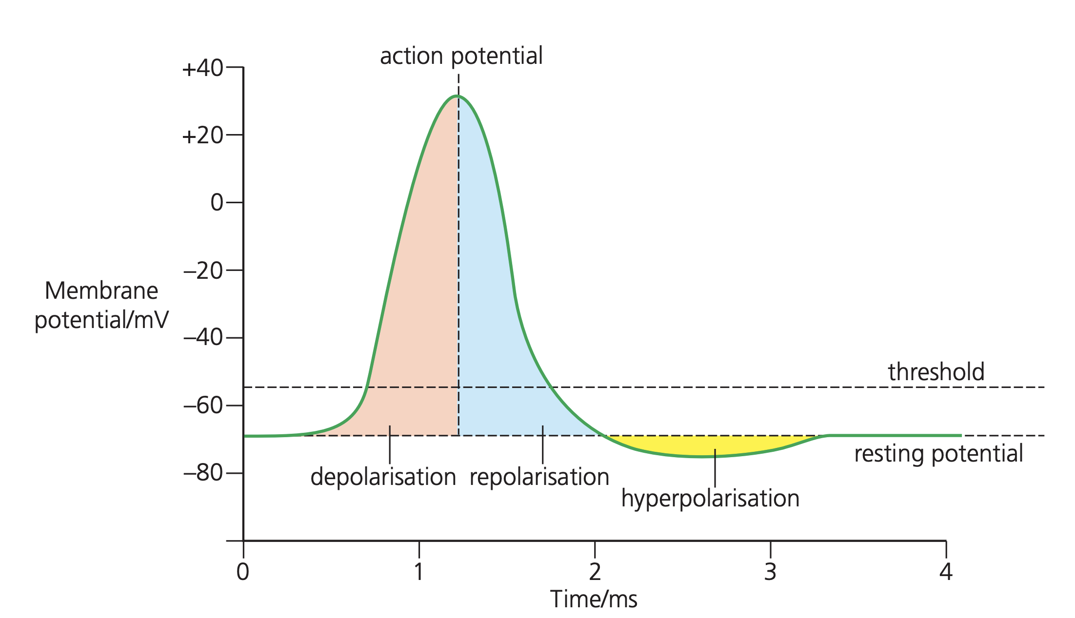
Measuring action potentials
Action potentials can be measured using electrodes connected to an oscilloscope, which displays the voltage changes over time. The trace shows the characteristic spike shape, with rapid depolarisation followed by repolarisation and brief hyperpolarisation.
Action potentials are very brief events, typically lasting 1-2 milliseconds. The frequency of action potentials can vary - if maintained at a constant frequency, multiple action potentials can be calculated per second.
The oscilloscope trace provides valuable insights into the timing and magnitude of voltage changes, making it an essential tool for studying nerve impulse transmission.
Key Points to Remember:
- The resting potential (-65mV) is maintained by the sodium-potassium pump and selective membrane permeability
- Action potentials (+40mV) are triggered by sufficient stimuli and involve voltage-gated channel opening/closing
- The process follows six stages: rest → stimulus → sodium influx → peak → potassium efflux → repolarisation
- Voltage-gated channels respond to changes in membrane potential, creating positive feedback during depolarisation
- The entire action potential cycle takes only 1-2 milliseconds but allows rapid nerve signal transmission