Physics of the Eye (AQA A-Level Physics): Revision Notes
Physics of vision
Introduction to the eye as an optical system
The human eye operates as a sophisticated optical system, comparable to a video camera in its capabilities. This biological sensor contains approximately 130 million light-detecting cells and can function across an enormous range of light intensities—from near darkness to bright summer sunshine, where light levels may vary by a factor of 10 million.
The eye must meet three primary demands:
- Produce images in real time by updating the visual information approximately 20 times per second
- Automatically adjust focus from close-up objects (as near as 20 cm) to distant objects at infinity in less than one-tenth of a second
- Automatically adapt to changing light conditions
Both the eye and a video camera achieve these functions through similar mechanisms: they receive light through a variable aperture and use a convex lens to focus light onto a light-sensitive surface. A properly focused image occurs when light from each point on an object converges to a single corresponding point after passing through the optical system, creating a replica of the object.
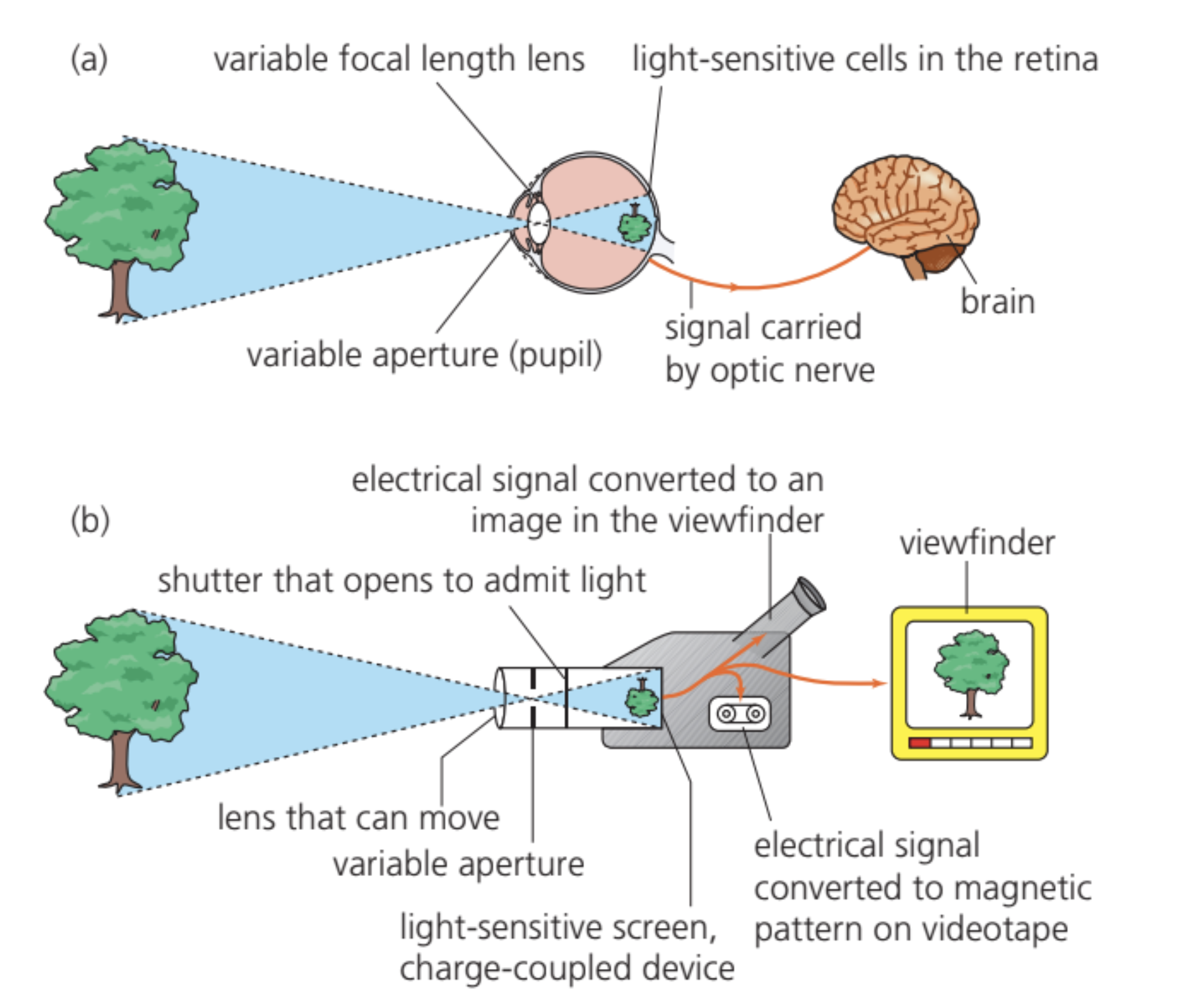
b) video camera-TV system
Controlling the amount of light entering the eye
The pupil serves as the eye's variable aperture, positioned in front of the lens. The diameter of the pupil is controlled by the iris, which consists of a ring of smooth muscle containing two types of muscle fibres. Some fibres run radially outward from the pupil like bicycle spokes, while others encircle the pupil. When the circular fibres contract, the pupil becomes smaller (constricts). When the radial fibres contract, the pupil opens wider (dilates).
In low light conditions, the pupil dilates to a maximum diameter of approximately 8 mm. In bright light conditions, it constricts to approximately 1.5 mm. This adjustment changes the area through which light enters the eye.
Pupil area calculation
Worked Example: Calculating Pupil Area Ratio
We can calculate the ratio of pupil areas between dilated and constricted states:
Area of constricted pupil:
Area of dilated pupil:
The ratio of these areas equals:
\text{Ratio} & = & \frac{\pi(4.0 \times 10^{-3})^2}{\pi(0.75 \times 10^{-3})^2} \\ & = & \left(\frac{4.0}{0.75}\right)^2 \\ & = & 28 \end{array}$$ This means the dilated pupil admits approximately :success[28 times more light] than the constricted pupil.Since the human eye can respond to light intensity changes by a factor of about one million, pupil size adjustment alone cannot account for the eye's full dynamic range. Other mechanisms, particularly those involving the retina's light-sensitive cells, play a more substantial role.
Forming a focused image through refraction
Light enters the eye through the cornea, a transparent membrane covering the front of the eye. The cornea is the first site where refraction occurs as light passes from air into the eye.
The amount of refraction at any boundary depends on the refractive index of the materials on either side of that boundary. The refractive index represents the ratio of the speed of light in air to its speed in the material.
Refractive Indices of Eye Components:
- Air:
- Cornea:
- Aqueous humour:
- Lens:
- Vitreous humour:
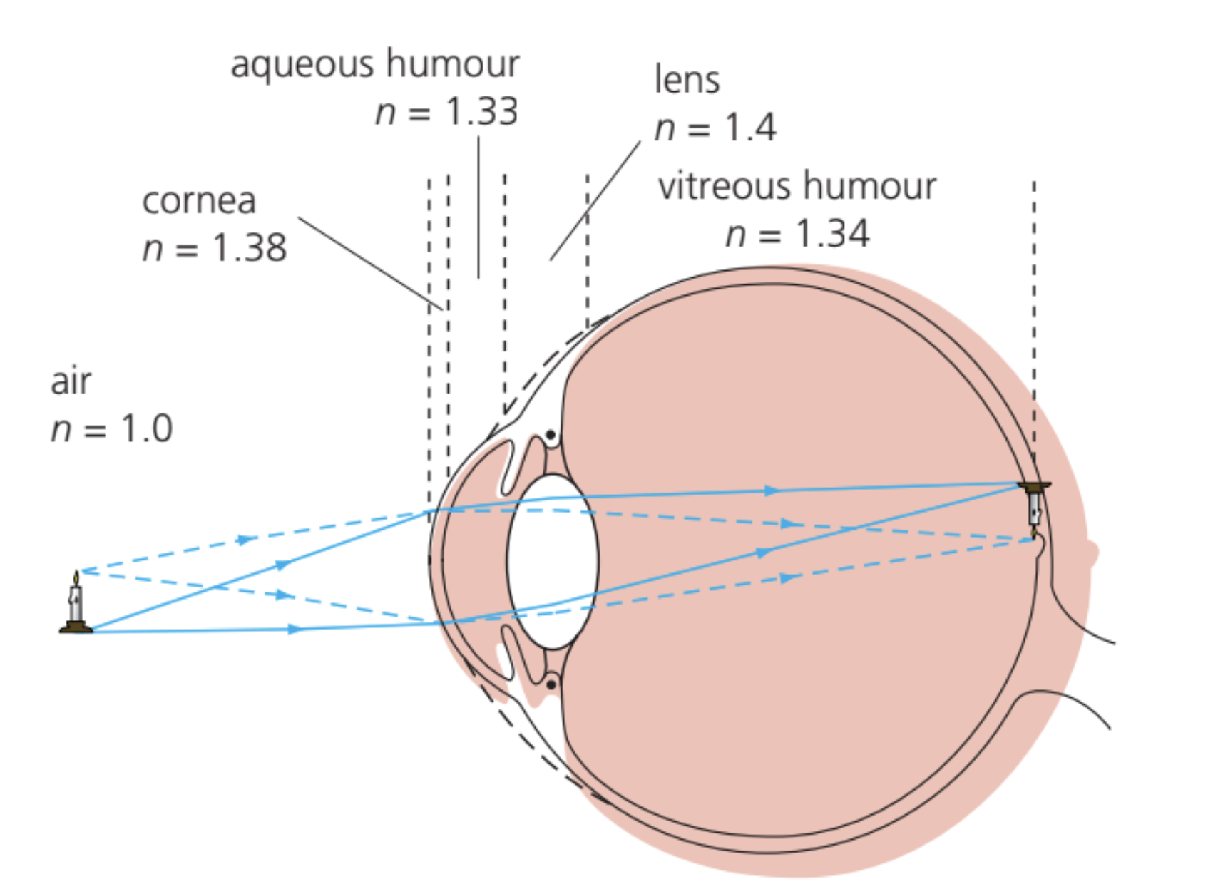
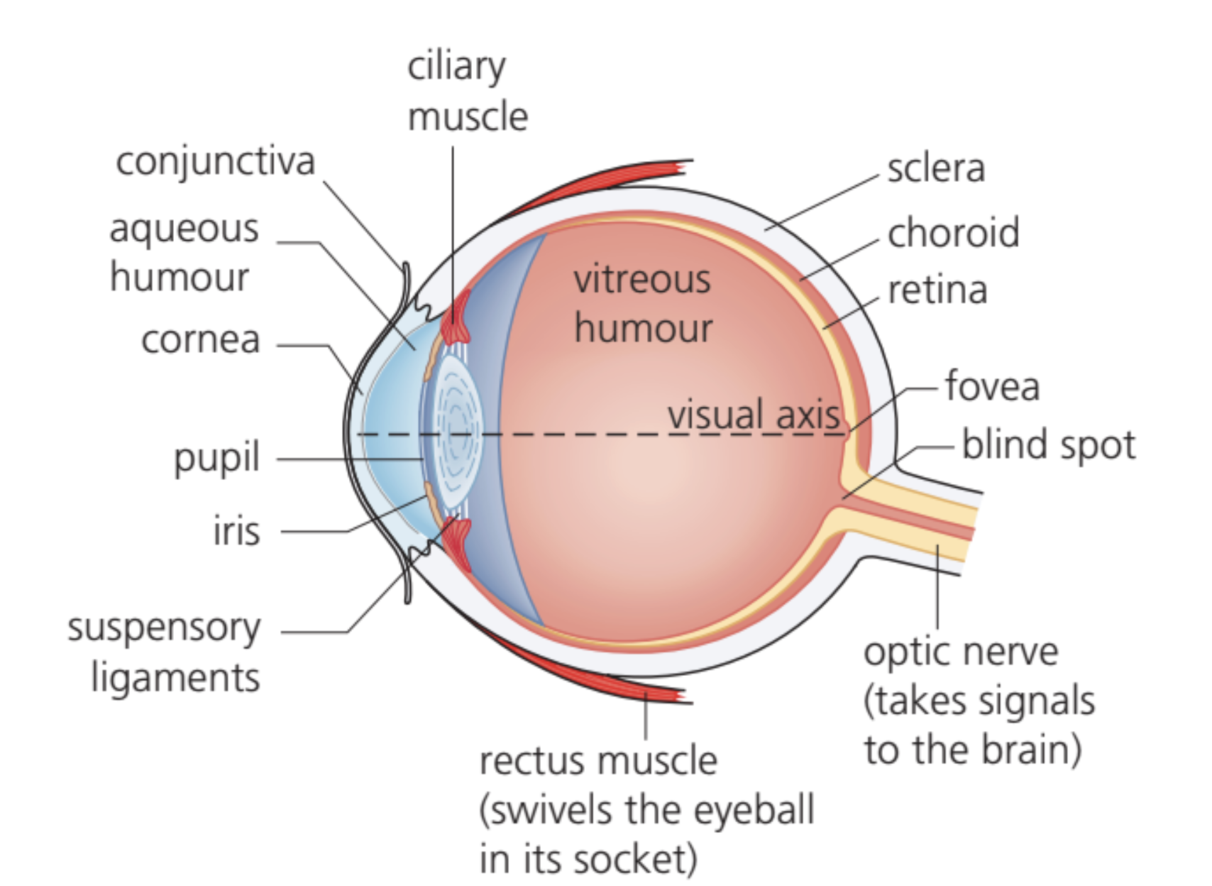
After passing through the cornea, light travels through the aqueous humour, a clear watery fluid, before reaching the lens. Light then passes through the lens and the vitreous humour (the gel-like substance filling most of the eyeball) before finally reaching the retina at the back of the eye.
At each boundary between these different media, light undergoes refraction. However, approximately 60% of the total refraction in the eye occurs at the air-cornea boundary. This is because the cornea presents a curved surface that bulges slightly outward from the eye, and the change in refractive index at this boundary (from 1.0 to 1.38) is the largest single change in the optical path. The second largest deviation occurs at the aqueous humour-lens boundary, which adds to the deviation caused at the cornea's front surface.
In an eye without defects, light from an object is focused to form a sharp image on the retina. The cornea's shape is particularly important—variations and defects in this shape can lead to focusing problems.
Sensitivity of the eye and the retina
The retina as a photodetector
The retina functions as the eye's photodetector, containing approximately 130 million light-sensitive cells. Each cell responds to light from one small point of the image. These cells contain chemicals called photopigments, which absorb photons of light and trigger the cell to generate electrical impulses. These electrical signals travel through a network of nerve fibers to the optic nerve and then to the brain for processing.
The retina contains two distinct types of light-sensitive cells:
Rod cells are extremely sensitive to light and can respond to very low illumination levels, even detecting individual photons. However, rods provide no information about colour. They are most sensitive to blue-green wavelengths around 500 nm. Rod cells contain a photopigment called rhodopsin, a light-sensitive protein.
Cone cells are less sensitive to light than rods but respond to narrower ranges of wavelengths. Their spectral response (response pattern according to wavelength) enables colour vision. Cone cells contain one of three different photopigments, each responding to different wavelength ranges.
Distribution of photoreceptors
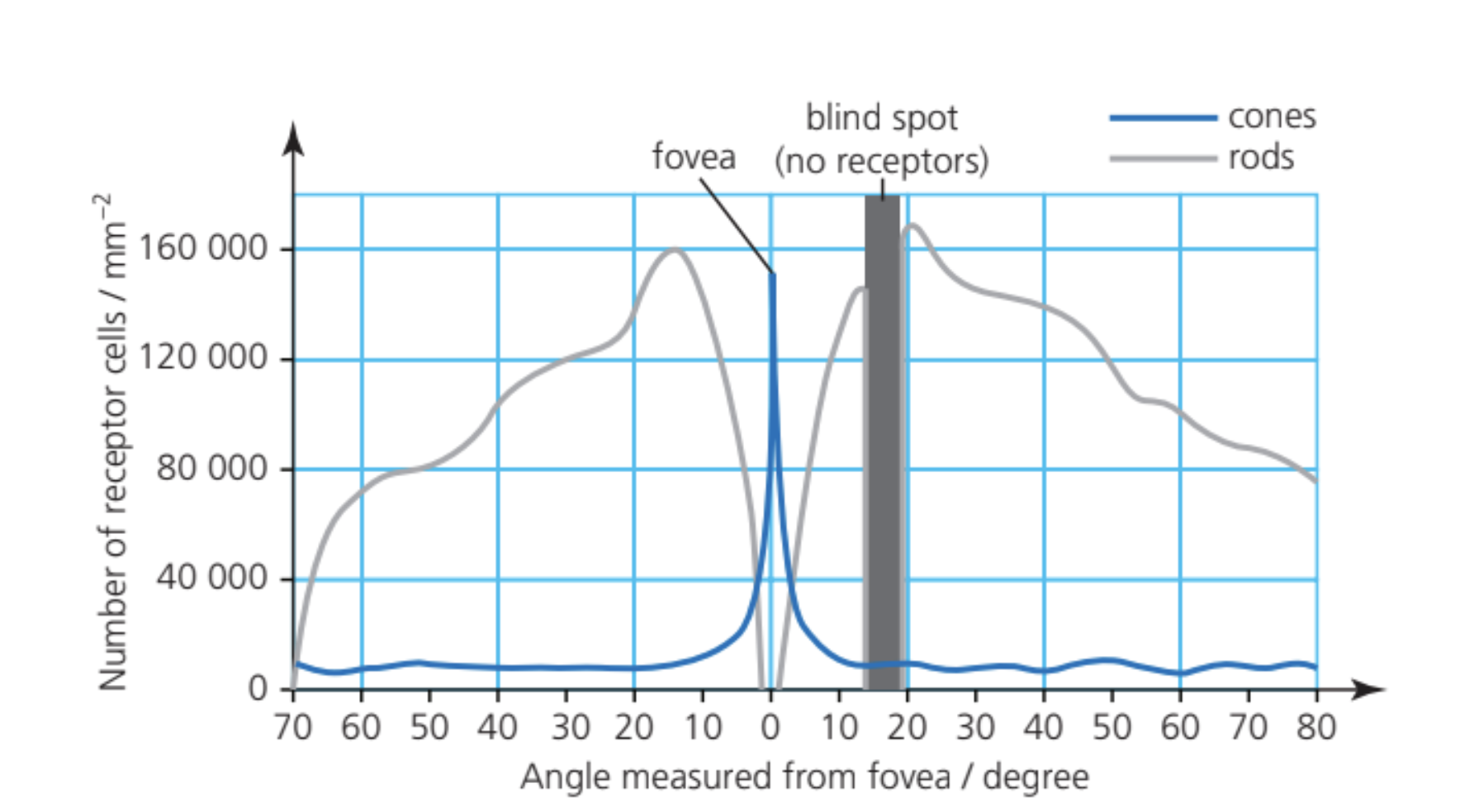
The density of rod and cone cells varies across the retina. At the center of the retina lies a small depression called the fovea, where cone density reaches its maximum—exceeding 150,000 cones per square millimetre. This region represents the center of our sharpest vision and the location of most colour perception. The fovea contains no rod cells and has no large blood vessels crossing it, with nerve fibers running radially away from it, providing an unobstructed path for light.
Toward the edges of the visual field, rod cells become more numerous than cones. The retina also has a blind spot, a region without any light receptors where the optic nerve exits the eye. Images falling on this region cannot be seen.
Photopic and scotopic vision
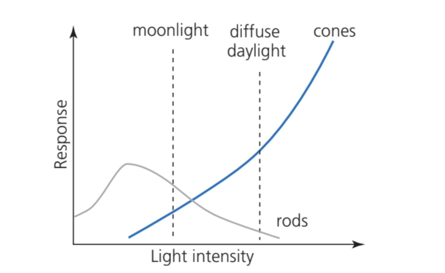
Under normal daylight conditions, vision relies primarily on cone cells. These conditions are called photopic conditions. Cones only function effectively in bright light.
In low light conditions, called scotopic conditions, rod cells become the primary photoreceptors. Different light intensities trigger different responses from these cells. In moonlight, rods dominate the visual response, while in diffuse daylight, both rods and cones contribute. In very bright conditions, cones provide most of the visual information.
Dark adaptation
When a photon is absorbed by rhodopsin in a rod cell, it triggers a small electrical current pulse lasting approximately 300 ms. During this period, the rod cannot detect any other photons. The absorption of light "bleaches" (destroys) rhodopsin molecules, and time is required for rhodopsin levels to recover.
In normal daylight, much of the rhodopsin remains bleached, so rods function at a low level and we rely on cones. When you move from bright sunlight into a darkened room, you initially cannot see clearly. There is insufficient light to stimulate cones, and the rods remain inactive due to bleached rhodopsin. However, as time passes, rhodopsin gradually regenerates and becomes active again, making your eyes increasingly sensitive to low light levels. This process is called dark adaptation.
Key Facts about Dark Adaptation:
- Full dark adaptation takes approximately 30 minutes
- When fully dark-adapted, rod cells can become up to 100,000 times more sensitive than in bright light
- This mechanism, rather than pupil size adjustment alone, accounts for the eye's remarkable ability to function across a wide range of light intensities
Colour vision
The eye can detect electromagnetic radiation over a wavelength range from approximately 380 nm to 750 nm. Rod cells respond across nearly this entire range, with peak sensitivity around 500 nm.
Cone Cell Types and Spectral Sensitivity:
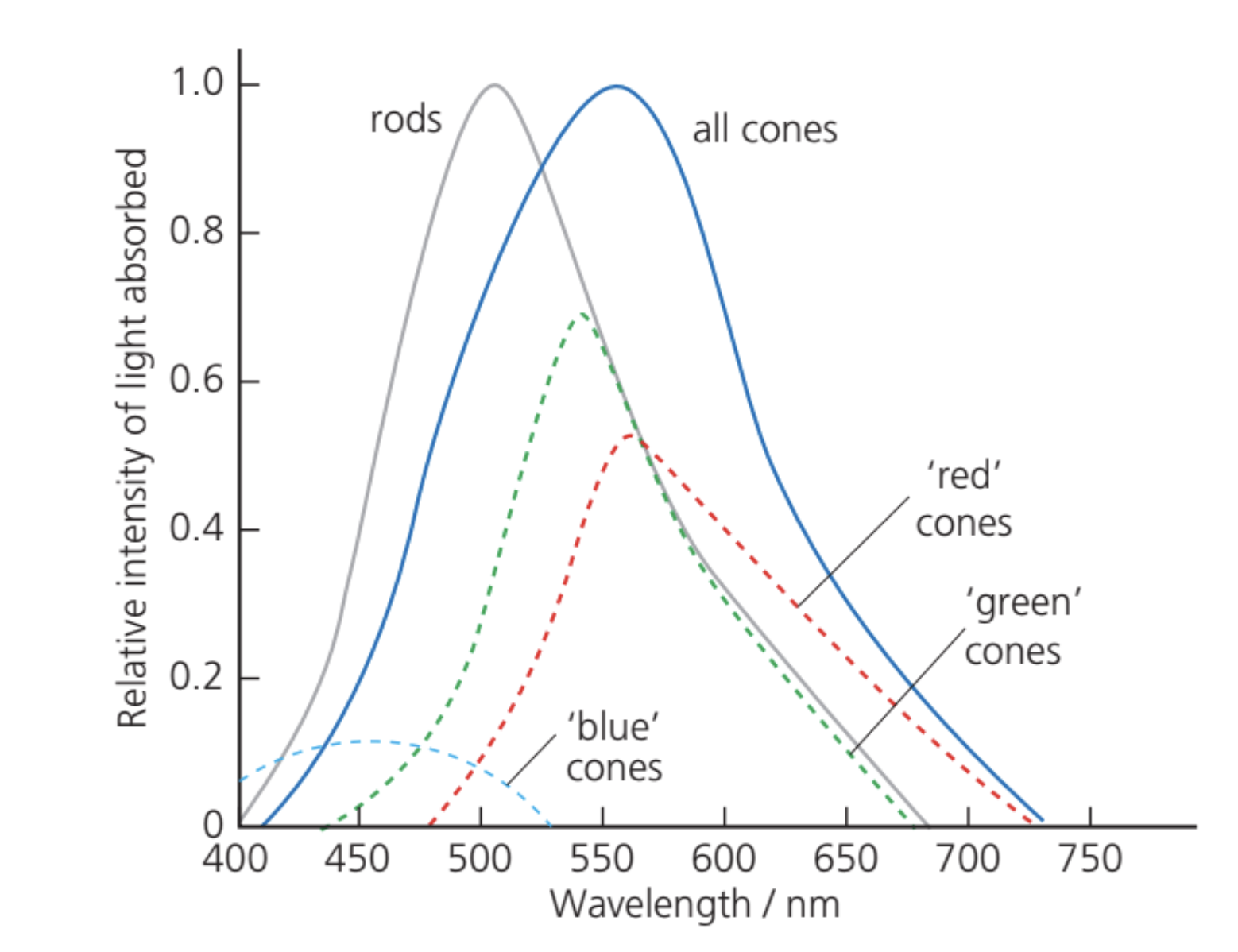
- 'Blue' cones: peak sensitivity around 420-450 nm
- 'Green' cones: peak sensitivity around 530-550 nm
- 'Red' cones: peak sensitivity around 560-580 nm
When a photon strikes the retina, one of these photopigments may absorb it. The probability of absorption depends on the photon's wavelength. Any given wavelength will stimulate a particular ratio of the three cone types. The brain interprets these ratios to identify wavelengths and create the sensation of colour.
Example: Perception of Yellow
The sensation of "yellow" results from stimulating both 'red' and 'green' cones in the appropriate proportion. Interestingly, yellow light on a television screen is actually produced by combining red and green light, which stimulates the same ratio of cone types as true yellow light would.
In very low light conditions, only rods function effectively. Since rods do not provide colour information, our ability to differentiate colours diminishes greatly in dim light. The range of wavelengths we can detect also narrows, as it becomes limited to the spectral response of rods rather than the combined range of all three cone types.
Spatial resolution and visual acuity
The eye's ability to see fine detail depends on the spacing of rod and cone cells in the retina. Two separate objects can only be distinguished if at least one unstimulated cell lies between the cells they stimulate. If adjacent cells are stimulated without an unstimulated cell between them, the two objects appear as one.
The average separation of light-sensitive cells on the retina measures 0.003 mm. The distance from the retina to the center of the eye lens along the principal axis is approximately 15 mm.
Worked Example: Calculating Minimum Resolvable Angle
Using the small-angle approximation (), we can calculate the minimum angle that can be resolved:
Visual acuity
The eye's ability to resolve detail is quantified as visual acuity, defined by:
An arcminute (arcmin) is a unit of angular measurement equal to 1/60th of one degree.
Worked Example: Converting Angular Resolution to Visual Acuity
To convert the angular resolution from radians to arcminutes:
Step 1: Convert radians to degrees
Therefore:
Step 2: Convert degrees to arcminutes
Step 3: Calculate visual acuity
Enhanced resolution at the fovea
The eye can actually achieve better resolution than this average value because rods and cones are not uniformly distributed. The fovea, with its exceptionally high cone density and absence of obstructing blood vessels and nerve fibers, provides maximum visual acuity of approximately 2 arcmin⁻¹, corresponding to a resolution of 0.5 arcmin.
Image resolution in bright light is superior to that in dim light because cone cells are smaller than rod cells, allowing finer detail to be resolved when cones dominate vision.
Key Points to Remember:
- The pupil (controlled by the iris) adjusts from 1.5 mm to 8 mm diameter, changing light admission by a factor of about 28, but the eye's total dynamic range is approximately one million times, indicating other mechanisms are more important for sensitivity adjustment.
- Approximately 60% of the eye's total refraction occurs at the air-cornea boundary due to the large change in refractive index (from 1.0 to 1.38) and the cornea's curved shape.
- Rod cells contain rhodopsin and function in low light (scotopic conditions), while cone cells enable colour vision and work best in bright light (photopic conditions). Dark adaptation takes about 30 minutes and can increase rod sensitivity by up to 100,000 times.
- Colour vision results from three types of cone cells with different spectral responses ('blue', 'green', and 'red'), and the brain interprets the ratio of stimulation among these types to perceive colour across wavelengths from 380 nm to 750 nm.
- Visual acuity (ability to resolve detail) equals 1 divided by the minimum resolvable angle in arcminutes. The fovea provides maximum visual acuity of about 2 arcmin⁻¹ due to its high cone density and unobstructed light path.