Electrical signals in the body (AQA A-Level Physics): Revision Notes
Electrical signals in the body
Introduction to bioelectrical signals
Electrical signals play a vital role in the human body, particularly in the nervous system and heart. These signals are generated by the movement of ions across cell membranes, creating changes in potential difference that can be measured and used for medical diagnosis.
Nerve cells and electrical conduction
Electrical signals travel through the body along specialised nerve cells. These nerve fibres can extend up to a metre in length while maintaining diameters of only a few micrometres. The signals propagate as changing potential differences generated by ion movement across the cell membrane.
Cell membrane properties
The nerve cell membrane exhibits selective permeability. Water molecules can diffuse freely through the membrane, but the membrane is much less permeable to sodium ions (Na⁺) and potassium ions (K⁺). However, specific channels exist within the membrane that allow these ions to transfer between the inside and outside of the cell.
The resting potential
Ion distribution at rest
In all cells at rest, there exists a characteristic ion distribution:
- Inside the cell: high potassium concentration, low sodium concentration
- Outside the cell: low potassium concentration, high sodium concentration
This unequal distribution of ions creates a potential difference across the cell membrane.
Establishing the resting potential
The concentration gradient causes potassium ions to diffuse out of the cell, taking their positive charges with them. As potassium ions leave, positive charge accumulates outside the cell. This process continues until the electrostatic force from the excess positive charge outside becomes strong enough to prevent further potassium ions from leaving.
Resting potential is the equilibrium potential difference across the cell membrane when the cell is at rest. At this point, the inside of the cell reaches a potential of −70 mV relative to its surroundings.
The cell is described as polarised when it maintains this resting potential. The polarised state represents an equilibrium where the concentration gradient (favouring ion movement) balances the potential gradient (opposing further ion movement).
The action potential in nerve cells
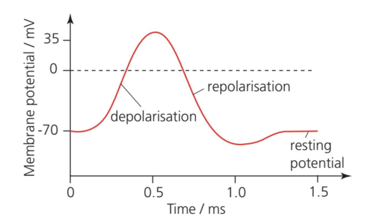
An action potential is the characteristic pattern of changing electrical potential that occurs when a nerve cell is stimulated. This changing potential allows signals to travel along nerve fibres.
Depolarisation
When a nerve impulse initiates, sodium channels in the cell membrane suddenly become much more permeable. Sodium ions rapidly flow into the cell. This process, lasting approximately 1 millisecond, causes the potential difference across the membrane to rise from −70 mV to +35 mV.
The cell is now depolarised, meaning the inside of the membrane has become positive relative to the outside, reversing the usual polarity.
Repolarisation
When the membrane potential reaches +35 mV, this positive potential triggers two effects:
- Sodium channels close, stopping the influx of sodium ions
- Potassium channels open, allowing potassium ions to leave the cell
As potassium ions flow out of the cell, they carry positive charges with them. This restores the resting potential of −70 mV. The cell is now repolarised, meaning it has returned to its original polarised state.
Restoration and propagation
After repolarisation, the original ion concentrations are gradually restored, returning sodium ions outside and potassium ions inside the cell. This restoration is achieved by active transport mechanisms.
The action potential propagates along the nerve fibre because the changing potential in one region initiates a new action potential in the adjacent region. This allows the electrical signal to travel the entire length of the nerve fibre.
The complete action potential cycle in a nerve cell takes approximately 1.5 milliseconds, with depolarisation occurring over about 5 milliseconds of this time.
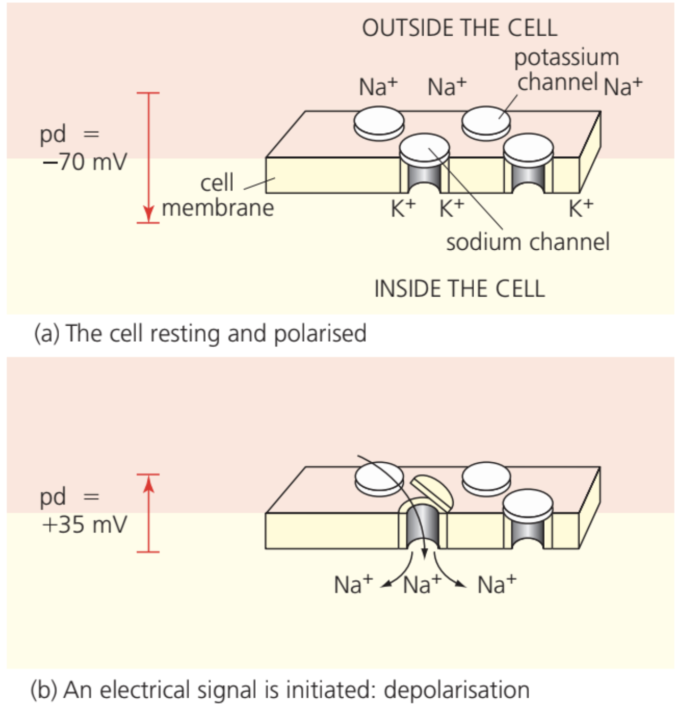
(a) In the resting state, there is an imbalance of ions, with more potassium ions inside the cell and more sodium ions outside. This creates a membrane potential of –70 mV, so the cell is polarised.
(b) Sodium channels open and sodium ions rush into the cell. The inside of the membrane becomes positive relative to the outside, meaning the membrane is depolarised.
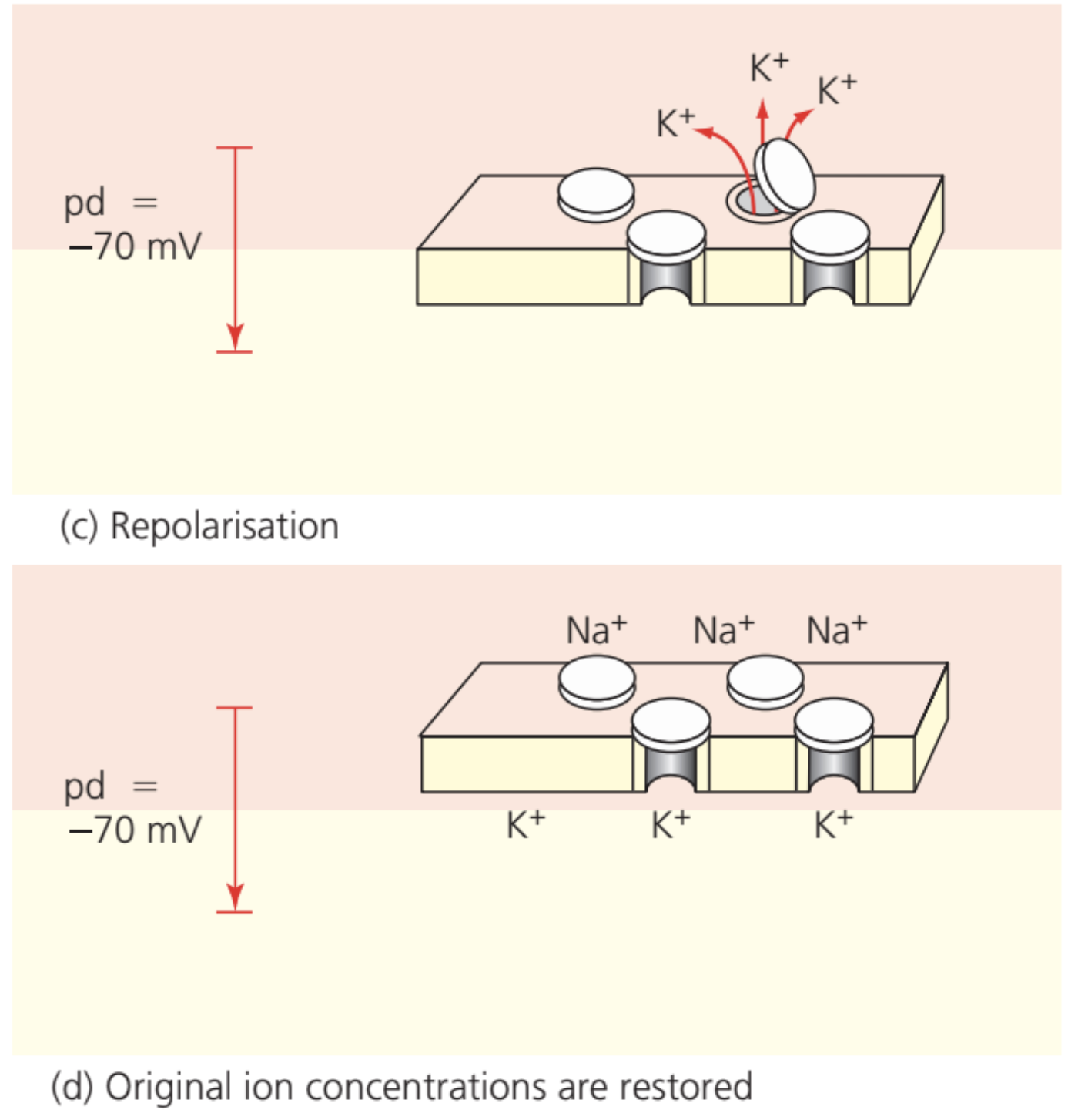
(c) The rise in membrane potential closes sodium channels and opens potassium channels. Potassium ions leave the cell, and the potential falls back to –70 mV; the membrane is repolarised.
(d) The original sodium and potassium ion concentrations are then restored.
Electrical activity in the heart
The cardiac conduction system
The heart contains a specialised structure called a node that functions as a natural pacemaker. Rather than being controlled by nerve cells, this node generates its own electrical impulses.
During each heartbeat, the electrical signal follows a specific pathway:
- The pacemaker node generates an electrical impulse
- The signal spreads rapidly across the atria (upper chambers), causing them to contract
- The signal then transfers to the ventricles (lower chambers)
- The ventricle walls contract uniformly, exerting pressure on blood from all sides
Action potential in heart muscle
The action potential in heart muscle shares similarities with that in nerve fibres but exhibits important differences:
Similarities:
- Same basic mechanism involving sodium and potassium ion movement
- Same sequence of depolarisation and repolarisation
- Same voltage changes (−70 mV resting, +35 mV depolarised)
Differences:
- The signal travels more slowly through heart muscle
- The entire cycle lasts substantially longer
- Depolarisation phase: approximately 0.2 seconds in heart muscle compared to 5 milliseconds in nerve cells
- The longer duration allows the heart muscle to relax completely before the next contraction begins
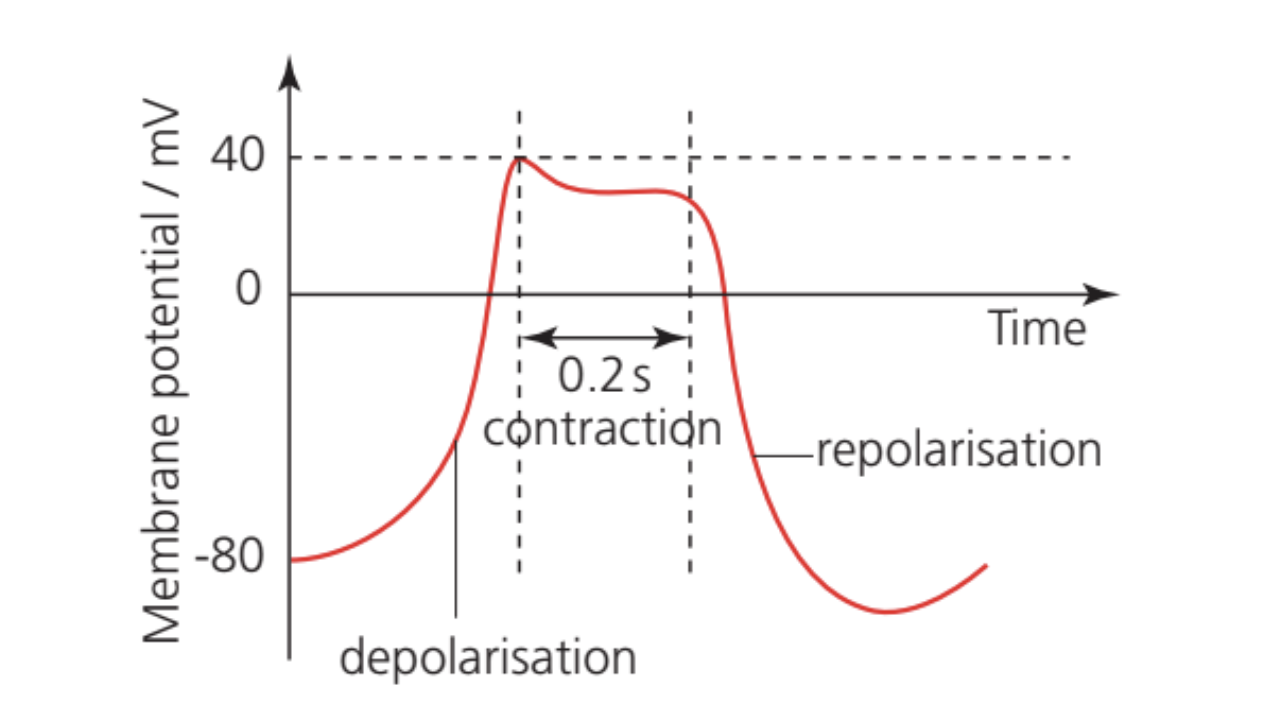
This extended action potential is necessary for proper heart function. The longer contraction phase ensures effective pumping of blood, while the extended relaxation phase allows the heart chambers to refill with blood before the next beat.
Relationship to the cardiac cycle
The electrical events correspond directly to mechanical events in the heart:
- Polarised cells → Heart muscle relaxed
- Depolarisation → Heart muscle contracts
- Repolarisation → Heart muscle relaxes
Remember!
Key Points to Remember:
- The resting potential of nerve and heart muscle cells is −70 mV, maintained by an imbalance of sodium and potassium ions across the membrane
- An action potential involves depolarisation (sodium ions enter, potential rises to +35 mV) followed by repolarisation (potassium ions leave, potential returns to −70 mV)
- Action potentials propagate along nerve fibres by triggering adjacent regions to depolarise
- Heart muscle action potentials last much longer than nerve action potentials (0.2 s vs 5 ms for depolarisation), allowing complete relaxation between beats
- The heart's natural pacemaker controls the electrical signals that coordinate atrial and ventricular contraction