X-ray Imaging (AQA A-Level Physics): Revision Notes
📚 Revision Notes
10.5.4 CT scanner
Introduction to CT Scanners:
- Traditional X-ray imaging generates 2D images, which only provide information about structures in one plane and lack depth.
- CT (Computed Tomography) scanners produce high-contrast images of cross-sections of the body, revealing depth and structure details that cannot be visualised with standard X-rays.
- By combining multiple cross-sectional images, a CT scanner can produce a 3D image of the area being investigated, giving a more complete view of the internal anatomy.
How a CT Scanner Works:
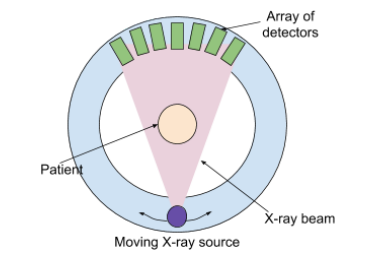
- Rotation of X-ray Tube: The X-ray tube revolves around the patient, emitting a narrow, monochromatic X-ray beam that passes through the body from various angles.
- Array of Detectors: Detectors are positioned around the patient's body, outside the path of the X-ray tube. Each detector measures the intensity of the X-ray beam after it has traversed the body.
- The detector directly opposite the X-ray source records the highest level of intensity as it passes through the least amount of tissue.
- Image Processing: The detectors send the recorded intensities to a computer, which compiles the data and constructs a cross-sectional image of the body.
Advantages of CT Scanning:
- Produces high-quality images of complex structures, such as bone fractures and soft tissues (e.g., brain).
- Non-invasive procedure with minimal physical discomfort for the patient.
- CT scanners yield a higher quality image than ultrasound and capture a complete cross-sectional view of the area.
Disadvantages of CT Scanning:
- CT scanning exposes patients to a higher dose of ionising radiation compared to standard X-rays.
- It is costly due to the complexity of the equipment.
- The contrast between materials with similar densities may be low, potentially causing image distortion.
- Patient movement can compromise image clarity, so patients are required to remain still, sometimes holding their breath, which can be challenging for certain individuals.
Understanding Image Quality and Contrast:
- CT scanners are excellent at differentiating between tissues of varying densities, making them ideal for detecting issues in areas like the brain and abdomen.
- However, materials with similar densities may be hard to distinguish from each other, potentially leading to lower contrast in the final image.
- Patient cooperation is essential for optimal results, as any movement can blur the image and reduce the accuracy of diagnosis.