The physics of diagnostic X-rays (AQA A-Level Physics): Revision Notes
10.5.1 The physics of diagnostic X-rays
Production of X-rays
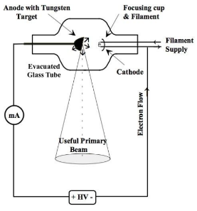
X-rays are produced through a process involving thermionic emission. This occurs when a metal is heated until the free electrons on its surface gain sufficient energy to escape. Here's how diagnostic X-rays are generated:
-
Electron Acceleration Electrons are emitted from a heated filament by thermionic emission in an evacuated tube and are then accelerated towards a metal target (anode) due to a high potential difference.
-
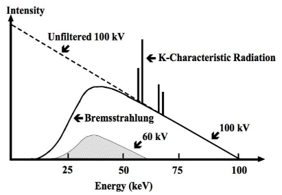
Collision and Energy Emission When these accelerated electrons collide with the metal target, they rapidly decelerate, releasing their energy in the form of electromagnetic (EM) radiation, specifically X-ray photons. This process is called bremsstrahlung or braking radiation, and it produces a continuous spectrum of X-rays.
-
Characteristic Radiation Some electrons will interact with the inner-shell electrons of the metal atoms, causing these inner electrons to be ejected. Outer-shell electrons then drop to lower energy levels to fill the vacancies, releasing energy in the form of characteristic X-ray photons. This emission forms a line spectrum superimposed on the continuous bremsstrahlung spectrum.
The maximum X-ray photon energy (and thus the X-ray spectrum's range) is determined by the product of the electron charge (e) and the accelerating voltage (V_a):
As the accelerating voltage increases, the total intensity of emitted X-rays increases, as does the peak photon energy. This results in higher energy X-rays and lower minimum wavelengths.
Efficiency of X-ray Production
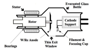
Only about 1% of the electron's kinetic energy is converted into X-rays; the rest raises the temperature of the target. Therefore, the target metal must be an efficient conductor of heat and have a high specific heat capacity. Tungsten is commonly used due to its high melting point and thermal conductivity.
To prevent overheating, the target/anode is often rotated at high speeds (around 3000 revolutions per minute) so the electron beam hits different areas, allowing for greater heat dissipation. The target is also typically designed with a bevelled edge to focus the electrons on a smaller area.
Intensity Control in X-rays
The intensity of the X-ray beam is defined as the total energy emitted per second per unit area. Two primary methods for controlling intensity are:
-
Increasing the Anode Voltage Higher anode voltage increases electron kinetic energy, leading to higher energy X-ray photons and increased intensity.
-
Increasing the Filament Current This increases the number of electrons emitted, hence more X-ray photons are produced per second. Here, only the intensity changes, not the energy of individual photons.
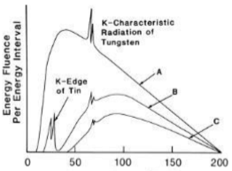
Image Sharpness and Radiation Safety
To enhance image sharpness:
-
The detection plate should be close to the patient.
-
Patients must hold still to avoid blurring.
-
A lead grid may be used to reduce scattered radiation that could reduce contrast. As X-rays are ionising radiation, exposure must be limited to avoid health risks. Key factors affecting radiation dose include:
-
Intensity of the X-ray beam.
-
Exposure time. Additionally, low-energy X-ray photons can be filtered out to reduce unnecessary dose to the skin, often using an aluminium philtre. Patients may also wear a lead-lined apron to protect other parts of their body from exposure.