Imaging techniques (AQA A-Level Physics): Revision Notes
📚 Revision Notes
10.6.1 Imaging techniques
Overview:
Radionuclide imaging involves using gamma-emitting radioisotopes as tracers to study specific areas of the body and assess how they function. These tracers are bound to substances that accumulate in targeted body parts. As these tracers travel through the body, they emit gamma radiation, which can be detected by a gamma camera to produce an image of the internal area of interest.
Key Radioisotopes Used as Tracers:
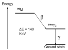
- Technetium-99m:
- Emits pure gamma radiation.
- Has a physical half-life of 6 hours, providing a balance between effectiveness and limited patient exposure.
- Easily prepared on-site and widely used due to its compatibility with various radiopharmaceuticals.
- Iodine-:
- Emits both beta and gamma radiation. Though beta emission is hazardous, iodine's natural uptake by the thyroid means it's particularly useful for thyroid investigations.
- Has a physical half-life of 8 days, providing enough time for detailed examination.
- Indium-:
- Also a pure gamma emitter with a half-life of 2.8 days.
- Often used in labelling antibodies and blood cells to detect infections, though it's more expensive than other tracers. | Radioisotope | Radiation Emitted | Physical Half-Life | Energy of Gamma Radiation | Primary Use | |---|---|---|---|---| | Technetium-m | Gamma | 6 hours | 140 keV | General use with radiopharmaceuticals | | Iodine- | Beta and gamma | 8 days | 360 keV | Thyroid imaging | | Indium- | Gamma | 2.8 days | 170 or 250 keV | Infection detection, cell labelling |
Production of Technetium-99m:
Due to its short half-life, Technetium-m cannot be transported over long distances. It is produced on-site using a Molybdenum-Technetium generator:
- Molybdenum-99 has a longer half-life of 66 hours, allowing easier transport to hospitals.
- The generator decays into Technetium-m, which can be isolated and injected into patients.
Positron Emission Tomography (PET)
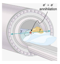
PET scanning uses positron-emitting radioisotopes for producing both 3D images and cross-sections of the body, primarily for assessing the metabolic activity of cells in a specific region. The steps include:
- Injection of the positron-emitting radioisotope attached to a substance localised to the target area.
- Absorption and decay of the radioisotope, releasing positrons that annihilate with electrons in the body.
- Annihilation produces gamma rays moving in opposite directions, which are detected and used to generate an image. The intensity of the image is related to metabolic activity:
- High metabolic activity (e.g., in tumours) results in greater radioisotope breakdown and more gamma emissions.
Advantages of PET Scans:
- Assess metabolic activity in regions of the body.
- Detect tumours and determine if they are spreading.
- Monitor brain activity, as gamma rays can penetrate the skull easily.
Disadvantages of PET Scans:
- Uses ionising radiation, which could damage cells.
- Scans take time, and patients must remain still, which may be uncomfortable or claustrophobic.
- PET scanners are expensive and less accessible, often requiring patients to travel to specialist facilities.