Gamma camera (AQA A-Level Physics): Revision Notes
10.6.3 Gamma camera
A gamma camera is an imaging device that detects gamma rays to create either 2D or 3D images (the latter if multiple cameras are used, as in a PET scanner). Gamma cameras are essential in medical imaging, particularly in nuclear medicine, as they allow doctors to visualise functional processes within the body.
How a Gamma Camera Works
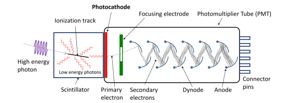
- Photomultiplier Tube: The core component of a gamma camera is the photomultiplier tube (PMT), which is an evacuated tube containing several parts:
- A photocathode that releases electrons when gamma photons hit it.
- Dynodes, which are electrodes set at increasingly higher positive potentials. Each dynode release more electrons when hit by the initial electrons, causing an electron multiplication process called secondary emission. This significantly amplifies the electron signal, ensuring that a measurable output signal is produced. Once the electrons have multiplied sufficiently, they reach the anode, where they create an electrical signal that can be processed.
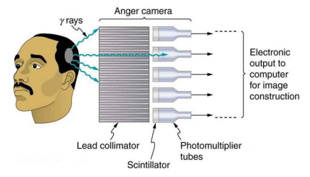
- Lead Collimator: Gamma rays entering the camera first pass through a lead collimator. This component is designed to philtre out scattered gamma photons by only allowing photons travelling parallel to the holes in the collimator to pass through. This helps to produce a sharper and more accurate image by reducing the background noise from scattered photons.
-
Scintillator: After passing through the collimator, the gamma photons reach the scintillator. The scintillator is a material that emits visible light photons when struck by gamma photons. This conversion from gamma radiation to visible light is crucial, as the photomultiplier tube can detect and amplify these light photons.
-
Image Creation: The light photons generated in the scintillator are captured by the photomultiplier tubes. The tubes then convert these light signals into electrical signals, which are processed by a computer. The computer reconstructs the spatial distribution of the gamma rays to form an image, which can be displayed on a monitor for diagnostic purposes.
Key Points
- Lead Collimator: Ensures that only gamma photons travelling parallel to the collimator holes are detected, improving image clarity.
- Scintillator: Converts gamma photons into visible light photons, which the photomultiplier tube can then process.
- Photomultiplier Tubes: Amplify the light signal through secondary electron emission, creating a signal strong enough to be processed into an image.
- Image Processing: A computer interprets the electrical signals from the photomultiplier tubes, constructing a visual image for medical analysis.
Application Example
In medical diagnostics, gamma cameras are used to assess organ function by detecting gamma radiation emitted by a radioactive tracer introduced into the patient's body. For example:
- In a thyroid scan, a radioactive iodine isotope is used. The gamma camera detects the radiation from the iodine to monitor thyroid function and identify areas with abnormal iodine uptake.