Neuronal Communication (OCR A-Level Biology A): Revision Notes
Neurones and Sensory Receptors
Communication and cell signalling
Multicellular organisms require coordination between different organs and systems. For effective functioning, cells must communicate with each other — a process called cell signalling. This communication allows the body to maintain homeostasis and respond to changes in the environment.
Both the nervous system and the hormonal system use cell signalling to transmit information. Signals can travel between cells that are close together or far apart. Only target cells respond to specific signals, ensuring precise control.
Example: Coordination During Exercise
During exercise, muscle tissue requires increased oxygen and glucose for respiration. The muscles themselves cannot supply these resources. Instead:
- Receptors detect changes (such as increased carbon dioxide or decreased oxygen)
- Signals are sent to the heart and breathing muscles
- The heart rate increases to boost circulation
- Breathing rate increases to enhance ventilation
This demonstrates how cell signalling coordinates different systems to meet the body's changing needs.
Structure of neurones
Neurones (nerve cells) are highly specialized cells that generate and transmit electrical impulses. Each neurone conducts impulses in only one direction. Bundles of neurones form nerves.
There are two main types of neurone based on the direction of impulse transmission:
- Sensory neurones carry impulses towards the central nervous system (CNS) from sensory receptors
- Motor neurones carry impulses away from the CNS to effectors
Remember: "Motor Moves Away" and "Sensory Sends In"
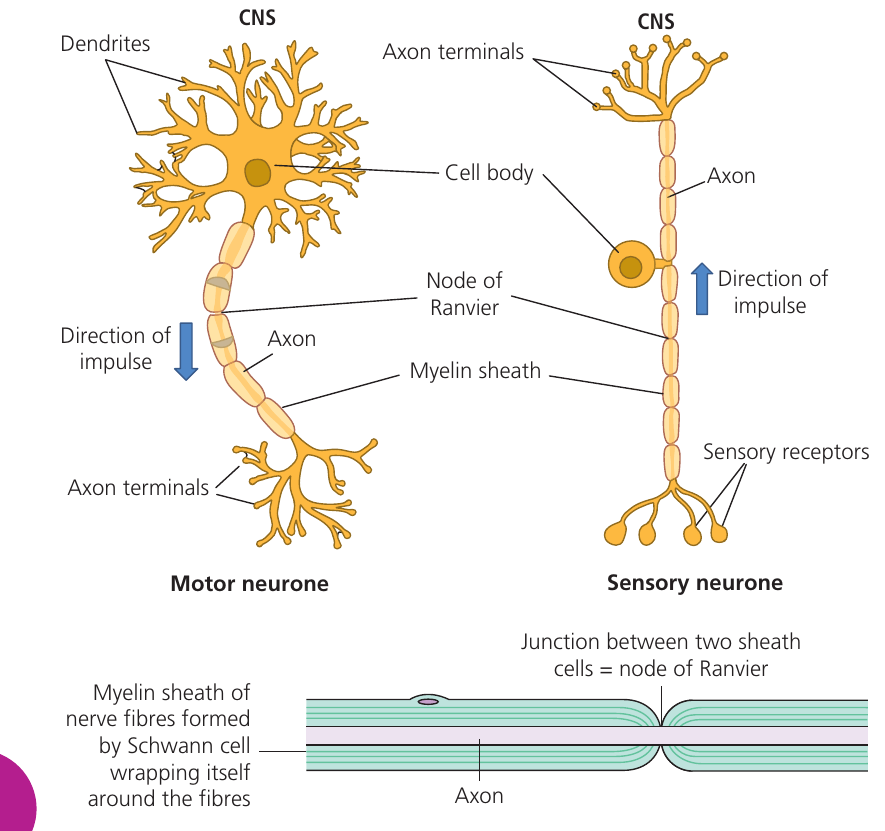
Key components of neurones
Both motor and sensory neurones share several structural features:
Cell body — contains the nucleus and organelles. In motor neurones, the cell body is positioned at one end of the cell and has branching processes called dendrites that connect to other neurones in the CNS. In sensory neurones, the cell body is located in the middle of the cell and lacks dendrites.
Axon — a long fiber that carries the nerve impulse. The axon can extend considerable distances to transmit signals across the body.
Myelin sheath — an insulating layer that surrounds the axon. Specialized cells called Schwann cells wrap themselves around the axon multiple times to form this sheath. The myelin is composed of lipid and protein, giving it electrical insulating properties.
The myelin sheath acts like the plastic insulation around an electrical wire, preventing the electrical impulse from dissipating and ensuring it travels efficiently along the axon.
Nodes of Ranvier — regular gaps in the myelin sheath along the length of the axon. At these points, the neurone's cell membrane is exposed and in direct contact with the extracellular fluid. These gaps play an important role in speeding up impulse transmission.
Axon terminals — branched endings at the end of the axon. In motor neurones, these transmit impulses to effectors (muscles or glands). In sensory neurones, they connect to other neurones in the CNS or directly to motor neurones in reflex arcs.
Comparison of motor and sensory neurones
| Feature | Motor neurone | Sensory neurone |
|---|---|---|
| Cell body position | At one end of the cell | In the middle (not at the end) |
| Dendrites | Present on cell body | Absent |
| Direction of impulse | Away from CNS to effector | Towards CNS from sensory receptor |
Other types of neurone
Relay neurones (also called interneurones) are found in the CNS. These cells have short axons and transmit signals from one neurone to another within the brain or spinal cord.
The neurones described above are myelinated — their axons have a myelin sheath. However, some neurones in the nervous system are non-myelinated and have bare axons.
The myelin sheath increases the speed of impulse transmission, but when impulses only travel very short distances (as often occurs in the CNS), myelination provides no advantage. This is why many relay neurones in the brain and spinal cord are non-myelinated.
Effectors
An effector is an organ that becomes active in response to a nerve impulse. In mammals, there are two types of effector:
- Muscles — contract when stimulated
- Glands — secrete substances when stimulated
Sensory receptors
Sensory receptors detect changes in the environment (stimuli) and convert them into electrical nerve impulses. They function as transducers — structures that convert one form of energy into another.
The term "transducer" refers to any device or structure that changes one form of energy into another. In biology, sensory receptors are biological transducers that convert various forms of energy (light, chemical, mechanical) into electrical nerve impulses that the nervous system can process.
Types of sensory receptors
Sensory receptors are classified according to the type of stimulus they detect:
Seven Types of Sensory Receptors:
- Photoreceptors detect light (found in the retina of the eye)
- Chemoreceptors detect chemicals (found in the nose for smell, tongue for taste, carotid arteries to monitor blood carbon dioxide, hypothalamus to detect various blood chemicals)
- Mechanoreceptors detect mechanical strain or stretching (found in the ear for sound and movement, skin for pressure and touch, muscles for stretch detection)
- Proprioceptors detect body position
- Baroreceptors detect blood pressure (found in blood vessel walls)
- Osmoreceptors detect the concentration of body fluids (found in the hypothalamus)
- Nociceptors detect tissue damage, producing the sensation of pain
Mnemonic: "Please Call Me Pretty, Babe, Or Never" (Photoreceptors, Chemoreceptors, Mechanoreceptors, Proprioceptors, Baroreceptors, Osmoreceptors, Nociceptors)
Receptors are distributed throughout the body, allowing detection of both external stimuli (from the environment) and internal stimuli (from within the body).
Pacinian corpuscles
Pacinian corpuscles are mechanoreceptors found in the skin that detect pressure. They provide a detailed example of how a sensory receptor works.

Structure
A Pacinian corpuscle consists of:
- Concentric layers of fibrous connective tissue called lamellae, separated by gel-like material
- A naked (unmyelinated) axon ending of a sensory neurone at the center
- The rest of the sensory neurone axon is covered by a myelin sheath
The structure resembles an onion, with the sensory nerve ending surrounded by multiple protective layers.
Function
How a Pacinian Corpuscle Detects Pressure:
When pressure is applied to the skin:
Step 1: The pressure distorts the lamellae
Step 2: This distortion is transferred to the naked axon ending in the center
Step 3: The stretching of the axon ending causes ion channels in the membrane to open
Step 4: This initiates a nerve impulse in the sensory neurone
The frequency of nerve impulses generated relates to the intensity of the pressure applied. A stronger pressure causes more frequent impulses, allowing the brain to interpret not only the presence of pressure but also its intensity.
Additionally, the number of Pacinian corpuscles stimulated provides information about the size and distribution of the pressure. This allows you to determine, for example, both the size and relative weight of an object placed on your hand without looking at it.
Key Points to Remember:
-
Neurones are specialized cells that transmit electrical impulses in one direction only
-
Motor neurones carry impulses away from the CNS to effectors; sensory neurones carry impulses towards the CNS from receptors
-
The myelin sheath is an insulating layer made of lipid and protein, produced by Schwann cells wrapping around the axon
-
Nodes of Ranvier are gaps in the myelin sheath that help speed up impulse transmission
-
Sensory receptors act as transducers, converting different forms of energy (light, chemicals, pressure, etc.) into electrical nerve impulses
-
Pacinian corpuscles in the skin detect pressure through distortion of their layered lamellae structure, which opens ion channels in the sensory neurone ending