Synapses (OCR A-Level Biology A): Revision Notes
Synapses
Introduction to synapses
Nerve cells must communicate with each other to coordinate activities throughout the body. This communication occurs at specialised junctions called synapses. A synapse is a connection between neurones where nerve impulses are transmitted from one cell to another. These structures are essential for the nervous system to function effectively.
At a synapse, the nerve impulse must cross a small gap between cells. Unlike electrical transmission along an axon, transmission across a synapse is chemical in nature. The transmitting neurone releases a chemical messenger called a neurotransmitter, which then initiates an action potential in the receiving neurone. This chemical transmission allows for sophisticated control and integration of neural signals.
Individual neurones can have between and synapses connecting them to other nerve cells. This enormous number of connections creates an incredible level of complexity in neural communication, enabling the sophisticated processing capabilities of the nervous system.
Structure of a synapse
A synapse has several key structural components that enable chemical transmission between neurones:
The presynaptic knob (or presynaptic terminal) is the swollen end of the transmitting neurone's axon. This structure contains:
- Voltage-gated calcium channels in its membrane
- Membrane-bound vesicles filled with neurotransmitter molecules
- Mitochondria to provide ATP for active processes
The synaptic cleft is a tiny gap (approximately nm wide) that separates the presynaptic and postsynaptic membranes. Neurotransmitter molecules must diffuse across this space.
The postsynaptic membrane is the surface of the receiving neurone that faces the synaptic cleft. This membrane contains:
- Receptor proteins that bind specifically to neurotransmitter molecules
- Ligand-gated ion channels (often sodium channels) that open when neurotransmitter binds
- Voltage-gated sodium channels that propagate the action potential
The structural organisation of the synapse reflects its function: the presynaptic side is specialised for neurotransmitter release (with vesicles and calcium channels), while the postsynaptic side is specialised for receiving the signal (with receptors and ligand-gated channels).
Mechanism of synaptic transmission
Synaptic transmission follows a precise sequence of events that converts an electrical signal into a chemical signal and back to an electrical signal:
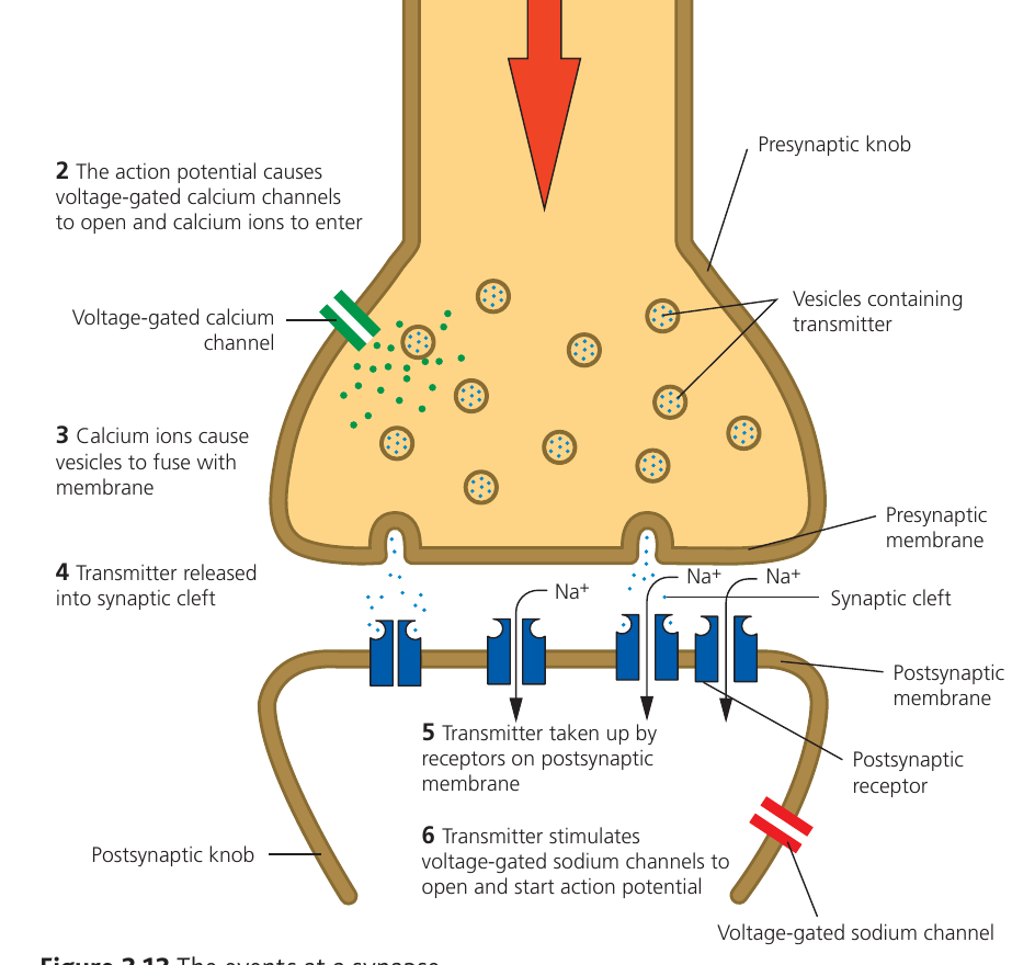
Step 1: Action potential arrival
When an action potential reaches the presynaptic knob, it causes depolarisation of the presynaptic membrane. The neurone terminal is separated from the postsynaptic membrane by the synaptic cleft.
Step 2: Opening of calcium channels
The depolarisation of the presynaptic membrane triggers voltage-gated calcium ion channels to open. These channels are specific proteins that respond to changes in membrane potential.
Step 3: Calcium influx and vesicle fusion
Calcium ion concentration is higher in the fluid outside the neurone than inside. When the channels open, calcium ions () diffuse into the presynaptic knob down their concentration gradient. The influx of calcium ions causes vesicles containing neurotransmitter to move towards the presynaptic membrane and fuse with it. The exact mechanism by which calcium ions trigger vesicle fusion remains an active area of research.
Step 4: Neurotransmitter release
Following vesicle fusion with the presynaptic membrane, the neurotransmitter is released into the synaptic cleft. The neurotransmitter molecules then diffuse across the synaptic cleft down a concentration gradient towards the postsynaptic membrane.
Step 5: Receptor binding
The postsynaptic membrane contains specific receptor proteins that have binding sites complementary to the neurotransmitter molecules. When neurotransmitter molecules reach the postsynaptic membrane, they bind to these receptors.
Step 6: Initiation of postsynaptic response
The binding of neurotransmitter to receptors acts as a chemical stimulus. This causes ligand-gated sodium ion channels to open in the postsynaptic membrane. If sufficient sodium ions () enter the postsynaptic neurone, the membrane potential reaches the threshold potential and a new action potential is generated.
Key Principle: Synaptic transmission involves converting an electrical signal (action potential) into a chemical signal (neurotransmitter release), and then back into an electrical signal (new action potential). This chemical step allows for signal modulation and control.
Neurotransmitters and cholinergic synapses
Many different chemicals can function as neurotransmitters in the nervous system. One of the most common and well-studied neurotransmitters is acetylcholine.
Cholinergic synapses are synapses that use acetylcholine as their neurotransmitter. These synapses are found throughout the nervous system, including at neuromuscular junctions where motor neurones meet muscle fibres.
After neurotransmitter release and binding, the neurotransmitter must be removed from the synaptic cleft. If acetylcholine remained in the synaptic cleft, it would continue to stimulate the postsynaptic neurone repeatedly (after each refractory period), even without new action potentials arriving from the presynaptic neurone.
Why Neurotransmitter Removal is Critical
To prevent continuous stimulation, cholinergic synapses contain an enzyme called cholinesterase (specifically acetylcholinesterase). This enzyme breaks down acetylcholine into smaller molecules. The breakdown products are then reabsorbed into the presynaptic knob by active transport, where they can be recycled to synthesise new acetylcholine molecules.
This removal and recycling process ensures that each action potential produces only one postsynaptic response, preventing uncontrolled nerve firing.
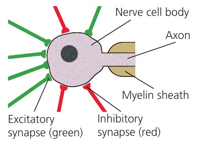
Types of synapses: excitatory and inhibitory
Synapses can be classified based on their effect on the postsynaptic neurone. The outcome depends on the interaction between the neurotransmitter and its receptor.
Excitatory synapses cause depolarisation of the postsynaptic membrane, making an action potential more likely to occur. These synapses work by opening sodium ion channels, allowing positive charge to enter the postsynaptic cell and bringing the membrane potential closer to the threshold potential. The neurotransmitters glutamic acid (the most common neurotransmitter in the central nervous system) and acetylcholine typically produce excitatory effects.
Inhibitory synapses make an action potential more difficult to achieve in the postsynaptic neurone. Instead of opening sodium channels, these synapses cause chloride ion channels to open. Chloride ions () carry a negative charge, so when they enter the postsynaptic neurone, the membrane potential becomes more negative (hyperpolarisation). This means a larger stimulus is required to reach the threshold potential. The neurotransmitter GABA (gamma-aminobutyric acid) is a common inhibitory neurotransmitter.
Distinguishing Excitatory from Inhibitory Synapses
- Excitatory synapses: Open channels → depolarisation → membrane potential moves closer to threshold → action potential more likely
- Inhibitory synapses: Open channels → hyperpolarisation → membrane potential moves further from threshold → action potential less likely
The presence of both types allows for sophisticated control of neural activity. A single neurone typically receives input from both types of synapse, and the final outcome depends on the balance between excitatory and inhibitory signals.
Role of synapses in the nervous system
Synapses enable complex communication and control within the nervous system. The fact that individual neurones can receive thousands of synaptic inputs creates enormous potential for integration and processing of information.
The combination of excitatory and inhibitory synapses provides subtle control over body functions.
Example: Breathing Control
Consider the example of breathing: breathing is normally an automatic reflex controlled by detecting blood carbon dioxide levels. When levels rise, excitatory signals are sent to the diaphragm and intercostal muscles to stimulate breathing movements.
However, there are situations when you might want to override this automatic response, such as when diving underwater. In this case, you can consciously activate inhibitory synapses that connect to the neurones controlling breathing muscles. The inhibitory input lowers the membrane potential of these neurones, making them less likely to fire. As a result, the normal excitatory signal from rising is insufficient to reach the threshold potential.
As carbon dioxide continues to accumulate in the blood, more excitatory synapses become activated and fire more frequently. Eventually, this increased excitatory input overcomes the inhibitory effect, and action potentials are generated to force breathing to resume.
This illustrates how the balance between excitatory and inhibitory inputs allows both automatic control and conscious override of neural responses.
Summation
The concept of summation is closely linked to the presence of multiple synapses on a single neurone. Summation refers to the way that signals from different synapses combine to determine whether the postsynaptic neurone will fire an action potential. The more impulses a neurone receives through its synapses, the more likely it is to reach threshold potential and generate an action potential.
There are two main types of summation:
Temporal summation occurs when a single synapse is activated repeatedly in quick succession. Each time the synapse fires, it releases neurotransmitter, which causes a small depolarisation of the postsynaptic membrane. If these signals arrive close enough together in time, the neurotransmitter accumulates in the synaptic cleft and the depolarisations add together. When the combined effect reaches threshold potential, an action potential is initiated in the postsynaptic neurone. The key feature is that one synapse fires multiple times.
Spatial summation occurs when several different synapses (all connected to the same postsynaptic neurone) transmit impulses at the same time. Each synapse contributes neurotransmitter and causes some depolarisation. The effects from these spatially separated synapses add together. If the combined depolarisation is sufficient to reach threshold potential, an action potential is generated. The key feature is that multiple synapses fire simultaneously.
Remembering the Difference:
- Temporal summation: "Temporal = Time" → one synapse firing repeatedly over time
- Spatial summation: "Spatial = Space" → multiple synapses at different locations firing together
Both types of summation demonstrate how the nervous system integrates multiple inputs to produce appropriate responses. They also allow for graded responses, where weak stimuli produce no action potential, moderate stimuli might trigger firing through summation, and strong stimuli reliably generate action potentials.
One-way transmission
The structure of synapses ensures that nerve impulses can only travel in one direction along a neural pathway. This unidirectional transmission is essential for organised information flow through the nervous system.
The structural basis for one-way transmission is clear: only the presynaptic side of the synapse contains vesicles filled with neurotransmitter, and only the postsynaptic side has receptors that can bind the neurotransmitter and ligand-gated channels that respond to it.
Why Backwards Transmission is Impossible
Because of this asymmetric arrangement, it is physically impossible for a nerve impulse to travel backwards across a synapse. The postsynaptic membrane cannot release neurotransmitter, and the presynaptic membrane lacks the receptors to respond to it.
This structural specialisation ensures that information flows in the correct direction through neural circuits, allowing for coordinated and purposeful responses to stimuli.
Remember!
Key Points to Remember:
-
Synapses are junctions between neurones where chemical transmission occurs via neurotransmitters such as acetylcholine
-
Synaptic transmission involves six key steps: action potential arrival → calcium channel opening → calcium influx → vesicle fusion → neurotransmitter release → receptor binding and postsynaptic response
-
Excitatory synapses (using neurotransmitters like acetylcholine or glutamic acid) cause depolarisation through sodium channel opening, while inhibitory synapses (using GABA) cause hyperpolarisation through chloride channel opening
-
Summation allows integration of multiple signals: temporal summation involves repeated firing of one synapse, while spatial summation involves simultaneous firing of multiple synapses
-
The asymmetric structure of synapses (vesicles only on presynaptic side, receptors only on postsynaptic side) ensures one-way transmission of nerve impulses