Mitochondria (OCR A-Level Biology A): Revision Notes
Mitochondria
What are mitochondria?
Mitochondria (singular: mitochondrion) are membrane-bound organelles located in the cytoplasm of eukaryotic cells. These rod-shaped structures typically measure in length, though some extend beyond this range, and in diameter.
The number of mitochondria within a cell correlates strongly with metabolic demand. Highly active cells such as liver hepatocytes and muscle fibres contain substantially more mitochondria than cells with lower energy requirements, such as adipocytes (fat-storage cells).
Mammalian red blood cells lack mitochondria entirely, as they rely solely on anaerobic respiration. This allows them to carry oxygen without consuming it for their own metabolic needs.
Mitochondrial structure
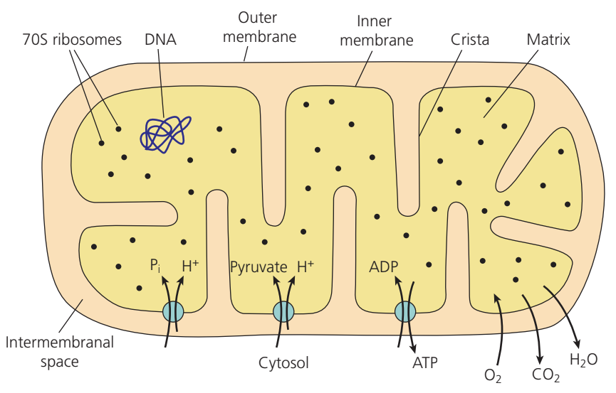
The structural organisation of mitochondria reflects their role in aerobic respiration and ATP synthesis. Each mitochondrion possesses several distinct compartments and components that work together to facilitate energy production.
The mitochondrial envelope
Mitochondria are enclosed by an envelope consisting of two separate phospholipid membrane layers. The outer membrane remains smooth, whilst the inner membrane exhibits extensive folding. Both membranes contain channel proteins, carrier molecules and various enzymes that regulate molecular transport and metabolic processes.
The two membranes differ in their phospholipid composition. This structural distinction provides supporting evidence for the theory of endosymbiosis, which proposes that mitochondria originated from ancient bacterial cells incorporated into early eukaryotic cells.
Cristae and surface area
The inner mitochondrial membrane folds extensively inward to form structures called cristae. These folds dramatically increase the available surface area, allowing the membrane to accommodate large numbers of electron carrier proteins and ATP synthase enzyme complexes. The greater the surface area, the more sites available for oxidative phosphorylation and ATP production.
The extensive folding pattern of cristae is directly related to the cell's energy needs. Cells with higher metabolic activity develop mitochondria with more densely packed cristae.
The intermembrane space
Between the outer and inner membranes lies the intermembrane space. This compartment plays a vital role in energy generation by maintaining a proton gradient. The space contains a higher concentration of hydrogen ions (H⁺) compared to both the cytosol and the matrix, resulting in a lower pH.
This concentration gradient represents potential energy – stored energy arising from the position of protons rather than their motion. When protons flow back through ATP synthase channels in the inner membrane, this potential energy converts into chemical energy stored in ATP molecules.
The mitochondrial matrix
Within the inner membrane, the matrix forms a gel-like region rich in proteins and lipids. This compartment houses several important components:
- A loop of mitochondrial DNA (mtDNA) – a circular, double-stranded DNA molecule resembling bacterial chromosomes
- 70S ribosomes – smaller ribosomes similar to those found in prokaryotes, distinct from the 80S ribosomes present in the eukaryotic cytoplasm
- Numerous enzyme molecules catalysing the link reaction, Krebs cycle, and contributing to the urea cycle
The matrix serves as the primary site for the link reaction and Krebs cycle during aerobic respiration. The presence of DNA and ribosomes enables the mitochondrion to synthesise some of its own proteins independently of the nuclear genome.
Structure, composition and function
The following table summarises the detailed organisation and roles of mitochondrial components:
| Structure | Composition | Function |
|---|---|---|
| Outer mitochondrial membrane | Phospholipid bilayer with embedded proteins | Permeable to pyruvate, oxygen (), carbon dioxide (), ATP and ADP, but impermeable to glucose |
| Inner mitochondrial membrane (folded into cristae) | Phospholipid bilayer containing protein complexes I–IV of the electron transport chain and ATP synthase | Protein complexes I, III and IV pump protons from the matrix to the intermembrane space; transport proteins facilitate movement of phosphate ions (), pyruvate and ADP from the intermembrane space into the matrix (movement of and pyruvate couples with inward proton flow); ADP exchanges for ATP; ATP synthase catalyses ATP synthesis from ADP and |
| Intermembrane space | Region with lower pH than both cytosol and matrix due to elevated proton (hydrogen ion) concentration | Maintains high proton concentration to drive proton movement through ATP synthase channels into the matrix |
| Matrix | Protein-rich gel containing a DNA loop, 70S ribosomes and numerous enzyme molecules | Site of the link reaction, Krebs cycle and part of the urea cycle; enables protein synthesis using mitochondrial ribosomes |
| Mitochondrial DNA (mtDNA) | Loop of double-stranded DNA | Codes for 13 proteins used within the mitochondrion; genes undergo transcription to produce mRNA; remaining mitochondrial proteins are encoded by nuclear DNA |
| 70S ribosomes | Composed of ribosomal RNA (rRNA) and proteins | Facilitate translation – the assembly of amino acids into polypeptide chains |
Evidence for endosymbiosis
The endosymbiosis theory proposes that mitochondria descended from free-living bacteria that became incorporated into ancestral eukaryotic cells. Several lines of evidence support this hypothesis:
Key Evidence Supporting Endosymbiosis:
- Mitochondria possess their own DNA arranged in a circular loop, mirroring the chromosome structure typical of bacteria
- The 70S ribosomes found in mitochondria match those present in prokaryotic cells, contrasting with the larger 80S ribosomes characteristic of eukaryotic cytoplasm
- The double membrane structure may represent the original bacterial membrane plus a membrane derived from the host cell during engulfment
- The different compositions of the outer and inner membranes suggest distinct evolutionary origins
Chloroplasts in plant cells show similar characteristics, indicating they too arose through endosymbiosis.
Adaptation to metabolic demand
Mitochondrial structure adapts to match cellular energy requirements. A trained athlete develops more mitochondria per cell, larger individual mitochondria, and increased numbers of cristae and ATP synthase complexes within each organelle compared to an inactive individual. These adaptations directly enhance the capacity for aerobic respiration and ATP generation.
An individual with moderate activity levels displays mitochondrial characteristics intermediate between trained athletes and inactive individuals. The degree of adaptation depends on both the intensity of physical activity and the duration of training.
Key Points to Remember:
- Mitochondria are rod-shaped organelles ( long, diameter) found in eukaryotic cells, with numbers varying according to metabolic demand
- The double membrane envelope consists of a smooth outer membrane and an extensively folded inner membrane forming cristae, which increase surface area for electron transport chains and ATP synthase
- The intermembrane space maintains a high proton concentration (low pH), creating potential energy that drives ATP synthesis when protons flow through ATP synthase into the matrix
- The matrix contains mitochondrial DNA (circular loop), 70S ribosomes, and enzymes for the link reaction and Krebs cycle, enabling partial independence from nuclear control
- Evidence supporting endosymbiosis includes bacterial-like DNA structure, 70S ribosomes, and distinct membrane compositions, suggesting mitochondria evolved from incorporated bacterial cells