Reflex Actions (OCR A-Level Biology A): Revision Notes
Reflex Actions
A reflex action is an automatic, involuntary behaviour triggered by a specific stimulus. These responses occur rapidly without conscious control and typically serve a protective function for the organism.
Characteristics of reflex actions
Reflex actions share three key features that distinguish them from voluntary movements:
- Involuntary – they occur automatically without conscious thought or decision-making
- Rapid – the response happens very quickly after the stimulus is detected
- Protective – they usually have survival value, protecting the body from harm
These three characteristics work together to make reflexes effective survival mechanisms. The rapid, automatic nature of reflexes means they can protect the body before conscious decision-making could even begin.
Examples of reflex actions
The human body performs many different reflex actions, including:
- Yawning
- Production of saliva
- Swallowing
- Withdrawal reflex (pulling a body part away from a source of pain)
- Blinking
- Pupil reflex (iris muscles constricting in bright light)
Some of these actions can also be performed voluntarily. For example, you can choose to blink. However, when carried out voluntarily, the action is no longer classified as a reflex because it involves conscious control.
The reflex arc
Every reflex follows a standard five-stage pathway called a reflex arc. The sequence always follows the same pattern:
Stimulus → Receptor → Coordinator → Effector → Response
Each component has a specific role in the reflex pathway:
- Stimulus – a detectable change in the environment that triggers the reflex
- Receptor – a sense organ or sensory cell that detects the stimulus
- Coordinator – the central nervous system (CNS), either the brain or spinal cord, which processes the signal
- Effector – the structure that carries out the response, usually a muscle but sometimes a gland
- Response – the action performed, which is always identical for a given stimulus
The response in a reflex arc is always the same for a specific stimulus. This predictability is a defining characteristic of reflex actions and distinguishes them from voluntary responses, which can vary.
These components are not unique to reflex actions. Voluntary actions also follow a similar sequence, but they involve conscious decision-making rather than occurring automatically.
The knee-jerk reflex
The knee-jerk reflex provides a clear example of a simple reflex pathway. Doctors commonly use this reflex to test that the nervous system is functioning correctly.
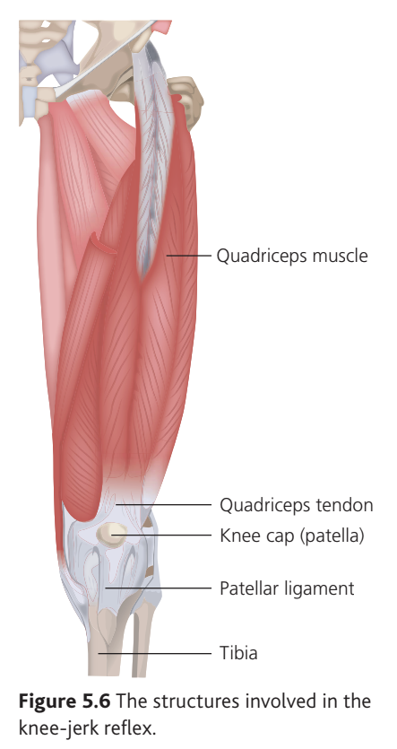
When you sit with your legs crossed and someone taps the patellar ligament (between the kneecap and tibia), your leg straightens involuntarily in a kicking motion.
The Knee-Jerk Reflex Pathway
The five components of this reflex are:
- Stimulus – stretching of the quadriceps muscle
- Receptor – stretch receptors within the quadriceps muscle
- Coordinator – the spinal cord
- Effector – the quadriceps muscle
- Response – contraction of the quadriceps muscle, causing the leg to straighten
Neural pathway of the knee-jerk reflex
When the patellar ligament is struck, it stretches the quadriceps muscle slightly. This stretching stimulates stretch receptors embedded in the muscle tissue. These receptors generate nerve impulses that travel along sensory neurones into the spinal cord.
Inside the spinal cord, the sensory neurone forms a synapse directly with a motor neurone. The impulse crosses this synapse and continues along the motor neurone to the quadriceps muscle (the effector). The muscle then contracts, causing the leg to straighten.
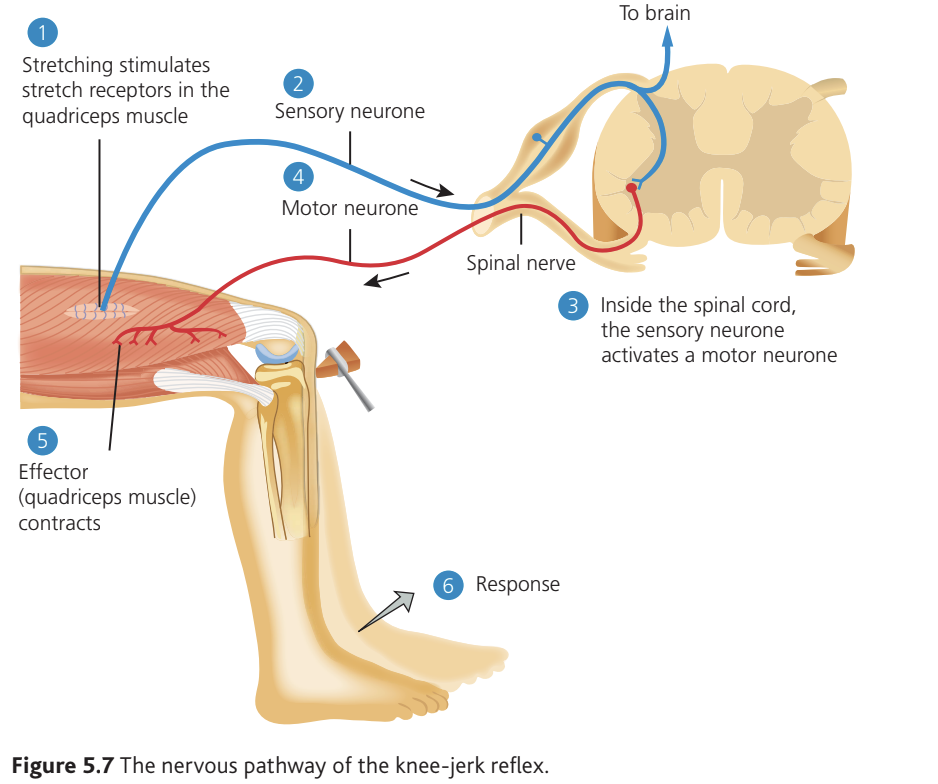
The knee-jerk reflex is unusual because there is no relay neurone involved. In most reflexes, a relay neurone in the CNS conveys impulses between the sensory and motor neurones. The knee-jerk reflex only involves one synapse.
In reality, multiple sensory neurones carry impulses from many stretch receptors, and multiple motor neurones stimulate different groups of muscle fibres. However, the basic pathway remains the same.
Why reflexes are rapid and automatic
The knee-jerk reflex pathway helps explain two key characteristics of all reflex actions: speed and automaticity.
Speed: Synapses create small delays in signal transmission. Each time a nerve impulse crosses a synapse, a brief pause occurs. The knee-jerk reflex only requires the signal to cross a single synapse between the sensory and motor neurones. If the signal had to travel through the brain, it would cross many more synapses, significantly slowing the response.
Automaticity: Connections within the spinal cord do send information about the stimulus to the brain. However, by the time the brain receives and processes this information, the response has already occurred. The brain therefore has no opportunity to make a conscious decision, and the response happens automatically.
Protective value of the knee-jerk reflex
The protective function of this reflex is not immediately obvious because it is usually triggered artificially during medical examination. However, the reflex actually forms part of the body's complex balance system.
When you start to fall forward, pressure on the patellar ligament stimulates the same stretch receptors in the quadriceps muscle. The resulting kicking action propels you upwards momentarily, giving your body time to adjust posture and prevent falling.
The blinking reflex
The blinking reflex protects the eyes from potential damage. It is more complex than the knee-jerk reflex because multiple effector muscles must work together to close the eyelids.
Stimuli that trigger blinking
Several different stimuli can initiate the blinking reflex:
- Anything contacting or irritating the cornea
- Drying of the cornea
- Objects rapidly approaching the eye
- Bright light
All of these stimuli could potentially damage the eye, making the rapid protective response valuable.
Neural pathway of the blinking reflex
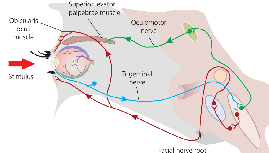
When the cornea is irritated or touched, sensory impulses travel along the trigeminal nerve (a cranial nerve carrying sensory information) to the medulla oblongata in the brain.
The reflex pathway passes through the brain this time, unlike the knee-jerk reflex which is coordinated entirely by the spinal cord. However, the signal does not reach the decision-making areas of the brain. Instead, it remains in the medulla where the number of synapses is kept to a minimum, ensuring the response remains rapid.
Effector muscles in the blinking reflex
Two main muscles work together to close the eyelids:
- Superior levator palpebrae muscle – lowers the upper eyelid
- Orbicularis oculi muscle – a circular muscle surrounding the eye that pulls the eyelids inwards and helps close them
Inside the medulla, the signal from the trigeminal nerve connects with multiple other neurones. Relay neurones are involved in the nervous pathway, particularly for controlling the lower eyelid. These relay neurones coordinate the signals sent to different motor neurones, which then stimulate the various effector muscles.
Despite being more complex than the knee-jerk reflex (due to multiple effectors and the presence of relay neurones), the blinking reflex still involves relatively few synapses. This keeps the response rapid and automatic, protecting the eyes before conscious thought can occur.
Key Points to Remember:
- Reflex actions are involuntary, rapid, and protective responses to stimuli that occur without conscious control.
- All reflexes follow the same sequence: stimulus → receptor → coordinator → effector → response.
- Reflexes are rapid because they involve minimal synapses – fewer synapses mean less delay in signal transmission.
- The knee-jerk reflex is coordinated by the spinal cord and involves only one synapse between sensory and motor neurones (no relay neurone).
- The blinking reflex is coordinated by the medulla and involves multiple effector muscles working together to close the eyelids and protect the eyes from damage.