Enzymes That Manipulate DNA (VCE SSCE Biology): Revision Notes
Enzymes That Manipulate DNA
Introduction to DNA manipulation enzymes
Scientists use specialised enzymes to manipulate DNA in various ways. These molecular tools allow us to cut, join, and copy DNA sequences with precision. Understanding how these enzymes work is essential for modern genetic engineering and biotechnology.
The three main types of enzymes used in DNA manipulation are:
- Endonucleases - cut DNA at specific sites
- Ligases - join DNA fragments together
- Polymerases - synthesise new DNA strands
Each enzyme plays a distinct role in DNA manipulation, much like the cut, paste, and copy functions on a computer.
Computer Analogy for DNA Manipulation
Just as computers use three fundamental operations (Ctrl+X to cut, Ctrl+V to paste, Ctrl+C to copy), scientists use three types of enzymes to manipulate DNA:
- Endonucleases = Cut (Ctrl+X)
- Ligases = Paste (Ctrl+V)
- Polymerases = Copy (Ctrl+C)
This analogy helps visualise how these molecular tools work together in genetic engineering.
Endonucleases - the molecular scissors
What are endonucleases?
Endonuclease is an enzyme that breaks the phosphodiester bond between two nucleotides in a polynucleotide chain. These enzymes act as molecular scissors, cutting DNA strands at specific locations.
When endonucleases target specific sequences, they are called restriction endonucleases (or restriction enzymes). These enzymes cut DNA by cleaving the phosphodiester bonds in the sugar-phosphate backbone that holds DNA nucleotides together. This cutting process is sometimes called "restriction endonuclease digestion".
Origins and naming of restriction endonucleases
Bacterial Origin of Restriction Enzymes
Restriction endonucleases are naturally found in bacteria, where they serve as a defence mechanism against invading viral DNA. When viruses inject their DNA into bacterial cells, restriction enzymes recognise and cut up the foreign DNA, protecting the bacteria from infection. Scientists have isolated these enzymes and repurposed them as powerful tools for DNA manipulation in laboratories.
The names of restriction endonucleases reflect the bacteria from which they were discovered. For example, EcoRI was discovered in the bacterium E. coli.
Recognition sites
A recognition site is a specific target sequence of DNA upon which restriction endonucleases act. These sites are typically four to six nucleotides long, and each enzyme recognises a unique sequence.
Palindromic Recognition Sequences
Recognition sites are usually palindromic sequences. This means the 5' to 3' sequence on one DNA strand is identical to the 5' to 3' sequence on the complementary strand.
For example, the enzyme TaqI recognises the sequence TCGA - reading from 5' to 3' on both strands gives the same sequence. This palindromic property is crucial because DNA is double-stranded, and the enzyme needs to recognise both strands to make its cut.
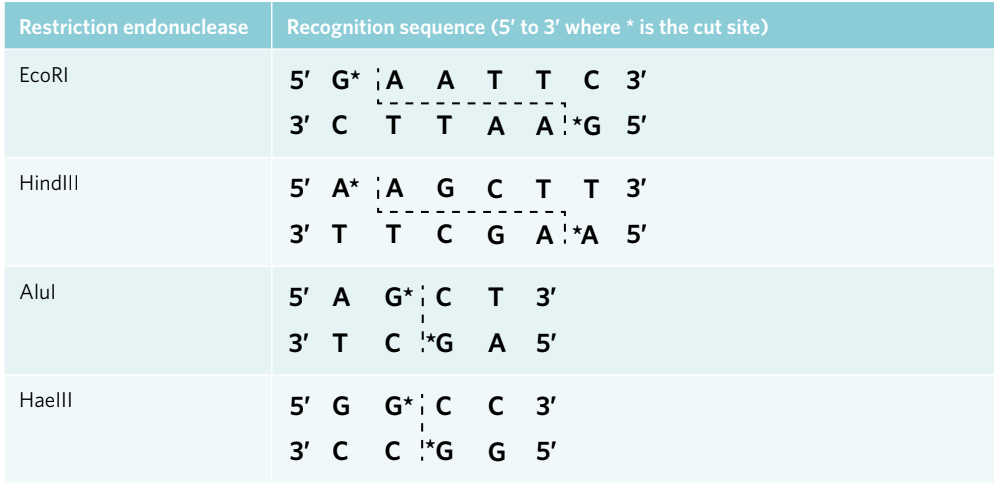
The table above shows recognition sequences for several common restriction endonucleases, with asterisks (*) marking where the enzyme cuts the DNA.
Sticky ends versus blunt ends
Restriction endonucleases create one of two types of cuts:
Blunt ends are the result of a straight cut across the double-stranded DNA by an endonuclease, resulting in no overhanging nucleotides. Enzymes like AluI and HaeIII cut straight through the middle of their recognition site, producing flush ends with no unpaired bases.
Sticky ends are the result of a staggered cut through double-stranded DNA by an endonuclease, resulting in overhanging nucleotides. Enzymes like EcoRI and HindIII make offset cuts that leave short, single-stranded regions. These overhanging nucleotides are unbonded nucleotides on the ends of the DNA strand resulting from a staggered cut.
Sticky ends are called "sticky" because the unpaired nucleotides are attracted to complementary sequences - they want to stick to matching bases through hydrogen bonding. This property is extremely useful in genetic engineering because it helps ensure that inserted genes are oriented correctly when joining DNA fragments.
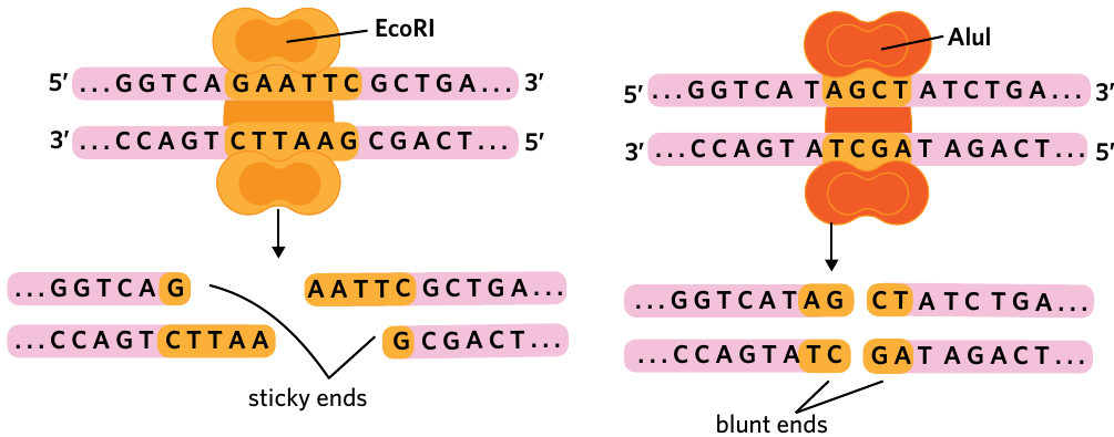
The diagram above illustrates how EcoRI creates sticky ends with overhanging nucleotides, while AluI produces blunt ends with no overhangs.
Cutting circular versus linear DNA
An important consideration when using restriction endonucleases is whether the DNA is circular (like plasmids) or linear.
For circular DNA, the number of fragments produced equals the number of recognition sites. If a plasmid has two recognition sites for a restriction enzyme, cutting at both sites will produce two DNA fragments.
For linear DNA, the number of fragments equals the number of recognition sites plus one. If a linear DNA molecule has two recognition sites, cutting at both will produce three fragments.
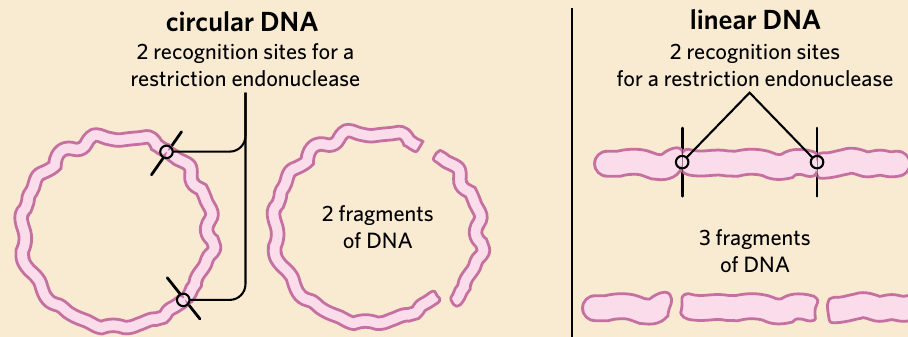
Worked Example: Calculating DNA Fragments
Question: A circular plasmid has 3 recognition sites for the restriction enzyme EcoRI. How many DNA fragments will be produced after complete digestion? What if the DNA were linear instead?
Solution:
Step 1: Apply the rule for circular DNA
- For circular DNA: number of fragments = number of cuts
- Number of cuts = 3
- Therefore: 3 fragments will be produced
Step 2: Apply the rule for linear DNA
- For linear DNA: number of fragments = number of cuts + 1
- Number of cuts = 3
- Therefore: 3 + 1 = 4 fragments will be produced
Key Rule to Remember:
- Circular DNA: n cuts = n fragments
- Linear DNA: n cuts = n+1 fragments
Exam Tip: Fragment Calculation Rule
This distinction between circular and linear DNA is crucial for exam questions:
- Circular DNA with n cuts = n fragments
- Linear DNA with n cuts = n+1 fragments
Think of it this way: Linear DNA already has two ends, so cutting creates additional fragments. Circular DNA has no ends, so each cut creates exactly one new fragment.
Ligases - the molecular glue
What are ligases?
Ligase is an enzyme that joins molecules, including DNA or RNA, together by catalysing the formation of phosphodiester bonds. While endonucleases cut DNA apart, ligases do the opposite - they join DNA fragments together, acting as molecular glue.
There are two main types of ligase enzymes:
- DNA ligase joins two DNA fragments
- RNA ligase joins two RNA fragments
How ligases work
Ligases join DNA fragments by creating phosphodiester bonds between the sugar-phosphate backbones of adjacent nucleotides. This process essentially reverses what endonucleases do.
Ligase Specificity
An important characteristic of ligases is their lack of specificity compared to restriction endonucleases. Ligases can join together any compatible blunt or sticky ends. This is because the substrates for ligase enzymes are the sugar and phosphate groups of DNA or RNA, rather than specific nitrogenous base sequences (which is the case for restriction endonucleases).
This versatility makes ligases incredibly useful in genetic engineering - they don't need to recognise specific sequences, they simply join any compatible ends together.
Joining sticky and blunt ends
Ligases can join both types of DNA ends:
Sticky end ligation: The complementary overhanging nucleotides on sticky ends base pair together first, holding the fragments in position. The ligase then forms phosphodiester bonds to permanently join the backbones. This process is more efficient than blunt end ligation because the base pairing helps hold the fragments in the correct orientation.
Blunt end ligation: Without overhanging nucleotides to hold fragments together, blunt end ligation is less efficient. However, ligases can still join blunt ends by bringing the flush ends close together and forming phosphodiester bonds.
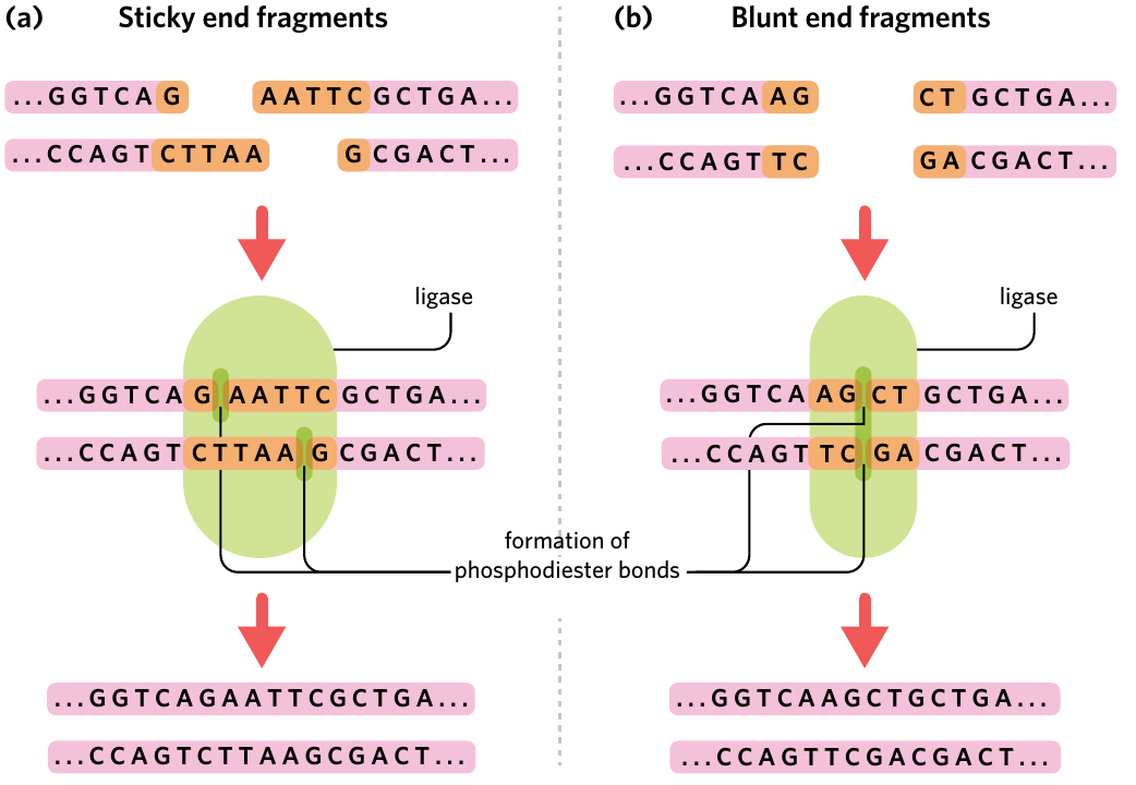
The diagram above shows how DNA ligase joins both sticky end fragments (part a) and blunt end fragments (part b) by catalysing the formation of phosphodiester bonds.
Key Difference: Sticky vs Blunt End Ligation
While ligases can join both types of ends, sticky end ligation is preferred in genetic engineering because:
- The overhanging nucleotides base pair together, holding fragments in position
- This base pairing ensures correct orientation of the inserted DNA
- The process is more efficient and reliable
- Blunt end ligation requires the fragments to be held together by chance, making it less predictable
Polymerases - the molecular copiers
What are polymerases?
Polymerase is an enzyme that synthesises a polymer from monomers, such as forming a DNA strand from nucleic acids. Polymerases build new strands of DNA or RNA by adding nucleotide building blocks one at a time.
Types of polymerases
The two main polymerases used in DNA manipulation are:
| Polymerase | Monomer | Polymer |
|---|---|---|
| RNA polymerase | RNA nucleotide | RNA strand |
| DNA polymerase | DNA nucleotide | DNA strand |
RNA polymerase is primarily used in transcription, where it synthesises RNA strands from DNA templates. This allows genes to be expressed by creating messenger RNA that ribosomes can translate into proteins.
DNA polymerase is used in DNA replication and amplification. This enzyme is particularly useful in forensic science, where investigators often have only tiny DNA samples. DNA polymerase can synthesise multiple copies of the DNA, providing enough material for analysis.
Forensic Application of DNA Polymerase
In forensic investigations, crime scene samples often contain only minuscule amounts of DNA - sometimes just a few cells from a hair follicle or a drop of blood. DNA polymerase enzymes can amplify these tiny samples through techniques like PCR (Polymerase Chain Reaction), creating millions of copies of the DNA. This amplification provides enough genetic material for detailed analysis and comparison, making it possible to identify suspects or victims from incredibly small samples.
How polymerases work
Polymerases require specific components to function:
- Template strand - A DNA strand that serves as the pattern for synthesis
- Primer - A short, single strand of nucleic acids that acts as a starting point for polymerase enzymes to attach
- Free nucleotides - The building blocks that will be added to create the new strand
The process works as follows:
- A primer binds to the template strand at the starting point
- The polymerase enzyme attaches to the primer
- The polymerase reads the template strand and adds complementary nucleotides
- The new strand is synthesised in the 5' to 3' direction
- The polymerase moves along the template, continuing to add nucleotides
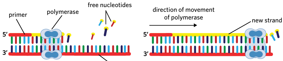
The diagram above illustrates DNA polymerase synthesising a new complementary strand from a template strand, with the primer marking the starting point.
Direction of synthesis
Directional Constraint of Polymerases
Polymerases can only synthesise DNA in the 5' to 3' direction. This means new nucleotides are always added to the 3' end of the growing strand. This directional constraint is crucial for understanding how DNA replication and synthesis occur.
This limitation is not arbitrary - it's determined by the chemistry of how phosphodiester bonds form. The enzyme can only catalyse bond formation between the 3'-OH group of the existing strand and the 5'-phosphate group of the incoming nucleotide.
Summary of DNA manipulation enzymes
The table below summarises the three main enzymes used in DNA manipulation:
| Enzyme | Action | Function |
|---|---|---|
| Restriction endonuclease | Cut | Cuts DNA or RNA at specific recognition sites |
| Ligase | Paste | Joins fragments of DNA or RNA together |
| Polymerase | Copy | Amplifies sections of DNA or RNA |
Key Characteristics to Remember
Endonucleases:
- Cut at specific recognition sites (usually 4-6 nucleotides)
- Recognition sites are typically palindromic
- Create either sticky ends (staggered cuts) or blunt ends (straight cuts)
- Named after bacteria they're found in
- Highly specific - each enzyme recognises one sequence
Ligases:
- Join DNA or RNA fragments by forming phosphodiester bonds
- Less specific than endonucleases
- Can join any compatible sticky or blunt ends
- Function as the reverse of endonucleases
Polymerases:
- Synthesise new DNA or RNA strands
- Require a primer to start synthesis
- Work in 5' to 3' direction only
- Use a template strand to determine nucleotide sequence
- Can amplify small DNA samples into larger quantities
Practical applications
These three enzymes work together in genetic engineering. For example, scientists have used these enzymes to insert a jellyfish gene for green fluorescent protein (GFP) into various organisms.
Worked Example: Inserting the GFP Gene
Scientists can make organisms glow by inserting the jellyfish gene for green fluorescent protein (GFP). Here's how the three enzymes work together:
Step 1: Cutting the gene Endonucleases cut the GFP gene from jellyfish DNA at specific recognition sites flanking the gene.
Step 2: Amplification Polymerases create multiple copies of the GFP gene, ensuring enough DNA material for the insertion process.
Step 3: Opening the recipient DNA Endonucleases cut open the recipient organism's DNA (such as a bacterial plasmid) at a specific recognition site.
Step 4: Insertion Ligases insert the GFP gene into the recipient's genome by joining the sticky or blunt ends together.
Result: The organism can now produce the fluorescent protein and glow under UV light! This technique has been successfully used in bacteria, plants, and even axolotl salamanders.
Remember!
-
Endonucleases are molecular scissors that cut DNA at specific recognition sites, which are usually palindromic sequences 4-6 nucleotides long
-
Sticky ends have overhanging nucleotides and can base pair with complementary sequences, while blunt ends are flush cuts with no overhangs
-
Ligases act as molecular glue, joining DNA fragments by forming phosphodiester bonds between sugar-phosphate backbones
-
Polymerases synthesise new DNA or RNA strands using a template and require a primer to begin synthesis in the 5' to 3' direction
-
For cutting DNA: circular molecules with n recognition sites produce n fragments, while linear molecules with n sites produce n+1 fragments