Exploring DNA Technology (VCE SSCE Biology): Revision Notes
Exploring DNA Technology
Introduction to DNA manipulation
DNA manipulation refers to a collection of laboratory techniques used by scientists to work with genetic material for various purposes. These techniques are essential tools in modern biology and have wide-ranging applications in biotechnology research, forensic investigations, and medical diagnostics. When scientists need to identify disease-causing mutations or solve criminal cases, they rely on these powerful methods to analyse DNA samples.
The three fundamental DNA manipulation techniques are:
-
DNA extraction - isolating DNA from cells
-
Polymerase chain reaction (PCR) - making multiple copies of DNA
-
Gel electrophoresis - separating DNA fragments by size
These techniques are often used in sequence, with each step preparing the DNA for the next stage of analysis. The output from one technique typically becomes the input for the next, creating a complete workflow for DNA analysis.
DNA extraction
What is DNA extraction?
DNA extraction is the process of isolating DNA molecules from cells. During this procedure, DNA is released from within the cell and separated from other cellular components such as organelles and proteins. This is a crucial first step in DNA analysis because scientists need pure DNA samples to work with.
Pure DNA samples are essential because contaminating proteins, lipids, and other cellular materials can interfere with subsequent analysis techniques like PCR and gel electrophoresis, leading to inaccurate or failed results.
To extract DNA, cells must first be broken open to release their contents. This process involves disrupting the cell membrane and nuclear envelope, which normally protect the DNA. Once the cells are broken open, various chemical treatments help to separate the DNA from proteins, lipids, and other cellular materials.
Key components of DNA extraction
The extraction process uses several important reagents:
Lysis solution: This solution contains two key components that break down cell structures. The first component disrupts cell membranes, whilst the second helps to break down proteins that are bound to the DNA. The lysis solution essentially acts to "unlock" the cells and release their DNA contents.
Salt solution: Salt is added to help the DNA precipitate out of solution. It neutralises the negative charges on the DNA molecules, allowing them to clump together and become visible.
Alcohol (usually ethanol or isopropanol): Cold alcohol causes the DNA to precipitate and form visible white strands that can be collected.
The extraction process
The main steps in DNA extraction include:
- Cell lysis - breaking open cells using lysis solution
- Protein removal - eliminating proteins that are attached to DNA
- DNA precipitation - using salt and alcohol to make DNA visible and collectible
- DNA washing - removing remaining impurities
- DNA resuspension - dissolving the purified DNA in a suitable buffer solution
Some steps involve using a centrifuge to spin down the sample, which separates DNA from other materials based on density. The sample may also be placed in a warm water bath to enhance the activity of enzymes that break down proteins.
Polymerase chain reaction (PCR)
What is PCR?
The polymerase chain reaction (PCR) is a technique that amplifies specific segments of DNA, creating millions of copies from a tiny initial sample. This amplification is essential because many DNA analysis methods require larger quantities of DNA than can typically be obtained from a biological sample.
PCR works by repeatedly copying a targeted region of DNA through cycles of heating and cooling. Each cycle doubles the amount of target DNA, so after just 30 cycles, a single DNA molecule can become over a billion copies.
The Power of Exponential Amplification
The exponential nature of PCR is remarkable. Starting with just one DNA molecule:
- After 10 cycles: 1,024 copies
- After 20 cycles: 1,048,576 copies
- After 30 cycles: over 1 billion copies
This makes PCR extremely sensitive and powerful for detecting even trace amounts of DNA.
The three stages of PCR
PCR operates through three distinct stages that occur in each cycle:
1. Denaturing (94-95°C): At this high temperature, the double-stranded DNA molecule separates into two single strands. The hydrogen bonds between complementary base pairs break apart, creating two template strands.
2. Annealing (50-56°C): As the temperature decreases, short DNA sequences called primers bind to complementary regions on each template strand. These primers mark the start and end points of the DNA region to be copied.
3. Extending (72°C): At this intermediate temperature, an enzyme called DNA polymerase adds nucleotides to the primers, building new complementary strands. This extends the primers and creates complete copies of the target DNA sequence.
Example: One Complete PCR Cycle
Starting material: One double-stranded DNA molecule
Step 1 - Denaturing (94-95°C):
- Heat breaks hydrogen bonds
- Two single-stranded templates formed
Step 2 - Annealing (50-56°C):
- Temperature drops
- Primers bind to complementary sequences on each strand
- Two primer-template complexes formed
Step 3 - Extending (72°C):
- Taq polymerase adds nucleotides
- New strands synthesized from primers
- Result: Two complete double-stranded DNA molecules
After one cycle: 1 DNA molecule becomes 2 molecules (doubled) After two cycles: 2 molecules become 4 molecules After three cycles: 4 molecules become 8 molecules
These three stages represent one cycle, and the entire process is typically repeated 25-35 times to produce sufficient copies of the target DNA.
Key components of PCR
Several essential components are needed for PCR to work:
Taq polymerase: This special DNA polymerase enzyme comes from a bacterium called Thermus aquaticus, which lives in hot springs. Unlike human DNA polymerase, Taq polymerase can withstand the high temperatures used in PCR without breaking down. This heat stability is crucial because the denaturing stage requires temperatures that would destroy most enzymes.
Why Taq Polymerase is Essential
Human DNA polymerase would be destroyed at the 94-95°C temperatures used for denaturing. Taq polymerase's ability to survive these extreme temperatures is what makes PCR possible. Without this heat-stable enzyme, the entire PCR process would fail after the first cycle.
Primers: These are short, single-stranded DNA sequences (usually 15-30 nucleotides long) that are complementary to sequences flanking the target DNA region. Primers are essential for specifying exactly which part of the genome will be amplified.
DNA template: This is the original DNA sample containing the sequence to be copied.
Nucleotides (dNTPs): These are the building blocks (adenine, thymine, guanine, and cytosine) that Taq polymerase uses to build new DNA strands.
Buffer solution: This maintains the optimal pH and provides necessary ions for the reaction.
Applications of PCR
PCR is widely used to amplify regions of interest within a genome, including both introns and exons. Introns are non-coding sequences that are removed during gene expression, whilst exons are the coding sequences that remain in the final messenger RNA and are translated into proteins. Being able to amplify specific regions allows scientists to study particular genes or identify genetic variations.
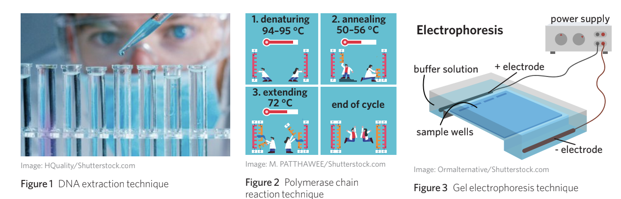
Gel electrophoresis
What is gel electrophoresis?
Gel electrophoresis is a technique used to separate DNA fragments according to their size and electrical charge. This method allows scientists to visualise and analyse DNA samples, making it possible to compare different samples or determine the sizes of DNA fragments present.
The technique works by applying an electric field to a gel matrix containing DNA samples. Because DNA molecules are negatively charged (due to the phosphate groups in their sugar-phosphate backbone), they migrate towards the positive electrode when an electric current is applied.
How gel electrophoresis works
The separation of DNA fragments occurs because smaller fragments can move through the gel matrix more easily than larger fragments. Think of it like trying to move through a crowded room - a small person can weave through gaps more quickly than a large person. Similarly, small DNA fragments travel further through the gel in a given time period, whilst large fragments move more slowly and remain closer to the starting point.
The Crowded Room Analogy
Imagine the gel as a crowded room full of obstacles. A child can quickly weave between people and reach the other side, while an adult carrying large bags moves much more slowly through the same space. This is exactly how DNA fragments behave in the gel - smaller fragments navigate the porous gel structure quickly, while larger fragments struggle to pass through.
This creates a pattern of bands on the gel, with each band representing DNA fragments of a similar size. The distance a band travels is inversely proportional to the size of the DNA fragments it contains - the further the band travels, the smaller the DNA fragments.
Key components of gel electrophoresis
Agarose gel: This is a porous matrix made by dissolving agarose powder in buffer solution and allowing it to set. The gel acts like a molecular sieve, creating a network of pores through which DNA molecules must navigate. The concentration of agarose determines the pore size - higher concentrations create smaller pores suitable for separating smaller DNA fragments.
Buffer solution: This liquid surrounds the gel and conducts electricity. It maintains a constant pH and provides ions necessary for electrical current to flow. Common buffers include TAE (Tris-acetate-EDTA) or TBE (Tris-borate-EDTA).
Sample wells: These are small indentations at one end of the gel where DNA samples are loaded before electrophoresis begins.
Power supply and electrodes: These create the electric field that drives DNA migration. The negative electrode (cathode) is placed at the end where samples are loaded, whilst the positive electrode (anode) is at the opposite end.
DNA standard (ladder): This is a mixture of DNA fragments of known sizes that is loaded into one well of the gel. It serves as a reference, allowing scientists to estimate the sizes of DNA fragments in unknown samples by comparing their migration distances to the standard. The DNA standard is typically loaded into the first well of the gel.
Visualising results
After electrophoresis is complete, the gel must be stained to make the DNA visible. Ethidium bromide is a commonly used fluorescent dye that intercalates (inserts) between the base pairs of DNA. When exposed to ultraviolet light, the dye fluoresces, making the DNA bands glow orange. This allows scientists to see and photograph the banding pattern.
Safety Consideration
Ethidium bromide is a powerful mutagen and must be handled with extreme care. Alternative stains like SYBR Green are increasingly used, particularly in teaching laboratories, as they are safer than ethidium bromide while still providing excellent visualization of DNA bands.
Reading gel electrophoresis results
The pattern of bands on a gel provides important information:
- Band position: Indicates the size of DNA fragments (closer to positive electrode = smaller fragments)
- Band intensity: Suggests the relative amount of DNA in that size range (brighter bands = more DNA)
- Number of bands: Shows how many different fragment sizes are present in the sample
By comparing sample bands to the DNA standard, scientists can determine the approximate size of DNA fragments in base pairs.
Applications of DNA technology
These three techniques work together in many real-world applications:
Forensic science: Crime scene investigators collect biological samples (such as hair, skin cells, or blood) from crime scenes. DNA is extracted from these samples, amplified using PCR, and then analysed using gel electrophoresis to create a DNA profile that can be compared to suspects or existing databases.
Medical diagnostics: Doctors can identify disease-causing mutations by extracting DNA from patient samples, amplifying the gene of interest using PCR, and analysing the results with gel electrophoresis. This can help diagnose genetic disorders or determine susceptibility to certain diseases.
Research: Scientists studying genetics use these techniques to investigate gene function, study evolutionary relationships between organisms, or develop new biotechnology applications such as genetically modified crops or gene therapy treatments.
The Complete DNA Technology Workflow
Notice how the three techniques form a complete analytical pipeline:
- Extract DNA from biological samples (DNA extraction)
- Amplify the specific DNA regions of interest (PCR)
- Analyse the amplified DNA fragments (Gel electrophoresis)
Each technique depends on the previous one, creating a powerful system for DNA analysis.
Key Points to Remember:
- DNA extraction isolates pure DNA from cells by breaking down cellular structures and removing proteins and other materials
- PCR amplifies specific DNA sequences through repeated cycles of heating and cooling, with three stages per cycle: denaturing (94-95°C), annealing (50-56°C), and extending (72°C)
- Taq polymerase is used in PCR because it can withstand high temperatures, unlike human DNA polymerase
- Gel electrophoresis separates DNA fragments by size using an electric field - smaller fragments travel further through the gel
- These three techniques are often used sequentially in forensic investigations, medical diagnostics, and biological research