Dissect a Sheep Heart (Leaving Cert Agricultural Science): Revision Notes
Dissect a Sheep Heart
Introduction
Heart dissection is a practical activity that allows you to examine the structure and function of one of the body's most important organs. A sheep heart is an excellent specimen for study as it's similar in size and structure to a human heart, making it perfect for understanding cardiovascular anatomy.
The sheep heart is particularly valuable for educational purposes because its size and structure closely match that of a human heart, while being more readily available for practical work.
Equipment required
For this dissection, you'll need several essential tools:
- Sheep heart - fresh or preserved specimen
- Dissecting board - provides a stable, hygienic work surface
- Scalpel - for making precise cuts through heart tissue
- Scissors - useful for trimming and detailed cutting
- Forceps - for holding and manipulating delicate structures
- Pins - to secure parts of the heart during examination
- Seeker - probe for exploring internal chambers and vessels
- Dropper - for adding fluids during the investigation
- Green food dye - to trace blood flow through coronary arteries
Preparation Tip: Ensure all equipment is clean and properly maintained before beginning. Sharp instruments work more effectively and are actually safer when properly maintained.
Heart anatomy overview
Before beginning the dissection, it's important to identify the external structures of the heart. The sheep heart shows several key features that are visible from the outside.
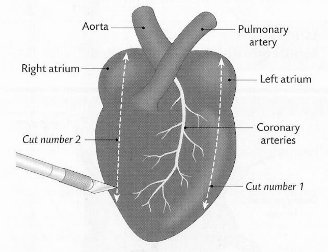
The major blood vessels extend from the top of the heart. The aorta is the largest artery, carrying oxygenated blood from the left ventricle to the body. The pulmonary artery carries deoxygenated blood from the right ventricle to the lungs. You can also identify the right atrium and left atrium, which are the upper chambers of the heart.
A distinctive feature visible on the heart's surface is the network of coronary arteries. These vessels appear as white, branching lines running diagonally across the front surface of the heart. They supply the heart muscle itself with oxygen and nutrients.
Identification Tip: The coronary arteries are your best landmark for orienting the heart correctly. They always run diagonally across the front (anterior) surface of the heart.
Step-by-step dissection method
Initial preparation and orientation
Start by placing the sheep heart on your dissecting board and take time to orient yourself with the specimen. Identify the front and back of the heart by locating the coronary arteries, which run diagonally across the front surface. This initial observation helps you understand the heart's natural position in the body.
Next, examine the major blood vessels connected to the heart. Look for the thick-walled aorta, the pulmonary artery, and the thinner-walled veins including the vena cava and pulmonary veins. Understanding these connections shows how blood flows to and from the heart.
Examining external features
Take note of any fat deposits surrounding the heart tissue. In living animals, this fat provides protection and insulation for the heart. The amount and distribution of fat can vary depending on the animal's age and condition.
Making the initial cuts
Step-by-Step Cutting Procedure
Step 1: Position your scalpel at the top of the heart
- Begin at the apex (pointed end) of the left ventricle
Step 2: Make the left ventricle cut
- Cut vertically down through the left ventricular wall
- Apply steady, controlled pressure
Step 3: Make the right ventricle cut
- Create a similar vertical cut through the right ventricle
- Keep cuts parallel for better examination access
Step 4: Open the chambers
- Gently spread the cut walls apart to expose internal structures
Exploring the ventricles
Once you've opened the ventricles, observe the thickness of the ventricular walls. You'll notice that the walls vary considerably in thickness. The left ventricle has much thicker walls than the right ventricle because it needs to pump blood at higher pressure to reach all parts of the body.
Look for the tricuspid valve (between right atrium and right ventricle) and bicuspid valve (between left atrium and left ventricle). These valves prevent blood from flowing backwards when the heart contracts.
Critical Observation: The difference in wall thickness between left and right ventricles is not a defect - it's a crucial adaptation! The left ventricle must generate enough pressure to pump blood throughout the entire body, while the right ventricle only needs to pump blood to the nearby lungs.
Examining the atria
Continue your dissection by cutting through the walls of the atria. Notice how much thinner the atrial walls are compared to the ventricular walls. This difference reflects their function - atria only need to push blood the short distance down into the ventricles.
Finding the heart valves
Identify the semi-lunar valves located at the base of the major arteries. These prevent blood from flowing back into the heart after it has been pumped out. The valve structure is designed to close when pressure decreases.
Valve Identification Guide
Tricuspid Valve: Located between right atrium and right ventricle
- Has three flaps (cusps)
- Prevents backflow during right ventricular contraction
Bicuspid (Mitral) Valve: Located between left atrium and left ventricle
- Has two flaps (cusps)
- Prevents backflow during left ventricular contraction
Semi-lunar Valves: Located at the base of aorta and pulmonary artery
- Crescent-shaped flaps
- Close to prevent arterial blood flowing back into ventricles
Studying the septum
Locate the septum, which is the muscular wall separating the left and right sides of the heart. Compare its thickness to the other heart walls. The septum is crucial for keeping oxygenated and deoxygenated blood separate.
Investigating coronary circulation
Find the opening to the coronary artery at the base of the aorta. Using your dropper, fill it with green food dye and gently pump the dye through the system. This demonstrates the pathway that supplies the heart muscle with blood and nutrients.
Coronary Circulation Investigation
Step 1: Locate the coronary artery opening
- Found at the base of the aorta, just above the semi-lunar valve
Step 2: Insert the dropper tip
- Gently place the dropper into the coronary artery opening
Step 3: Add green food dye
- Slowly squeeze the dropper to inject dye into the system
Step 4: Observer the dye pathway
- Watch as the dye travels through the coronary blood vessels
- Note how it branches across the heart surface
Key observations to record
Essential Observations to Document:
- Wall thickness differences between chambers and their functional significance
- Valve structure and position - how they prevent backflow
- Septum integrity - ensuring complete separation of heart sides
- Coronary artery distribution - how the heart supplies itself with blood
- Chamber size variations - relating structure to function
Safety considerations
Critical Safety Guidelines
- Always use sharp instruments carefully and cut away from your body
- Wear protective gloves when handling preserved specimens
- Work on a stable surface with good lighting
- Dispose of biological material according to school guidelines
- Wash hands thoroughly after the practical
Remember: Sharp instruments are safer than dull ones when used correctly - they require less pressure and give you better control.
Remember!
Key Learning Points:
- The sheep heart closely resembles human heart anatomy, making it an excellent model for study
- Left ventricular walls are thicker than right ventricular walls due to higher pressure requirements for systemic circulation
- Coronary arteries supply the heart muscle itself and can be traced using food dye techniques
- Four types of valves (tricuspid, bicuspid, and two semi-lunar valves) prevent blood backflow
- Systematic dissection reveals the relationship between heart structure and function, helping you understand how this vital organ works